If your neck or back feels tight, daily tasks like work, sleep, and exercise can become more difficult. This simple guide offers safe stretches to ease stiffness, improve posture, and help you move more comfortably. You'll learn when to stretch, how long to hold each stretch, and when it might be time to consult a clinician or physical therapist.
Before You Start: Safe Stretching Basics
Move slowly and stay in a comfortable stretch, not in pain. You should feel gentle tension that eases with steady breathing.
Hold most stretches 15 to 30 seconds, repeat 2 to 4 times per side, once or twice daily.
Stop and contact a clinician if you notice numbness, tingling, weakness, or pain that spreads into an arm or leg.
If you have osteoporosis, a recent injury, surgery, or active sciatica, ask your doctor or physical therapist which movements are best for you.
A Quick Daily Routine
Warm up with a short walk around the room for 1 to 2 minutes.
Do the neck sequence below, then the back sequence.
Finish with 3 slow breaths, in through your nose and out through your mouth, to help muscles relax.
Neck Stretches
These movements target common areas of tightness that can contribute to neck pain, tension headaches, and poor posture.
1) Chin Tucks
Sit or stand tall. Gently draw your chin straight back, as if making a small double chin. Keep your eyes level.
Hold 3 to 5 seconds, relax. Repeat 8 to 10 times.
Helps strengthen the deep neck flexors and counters forward head posture.
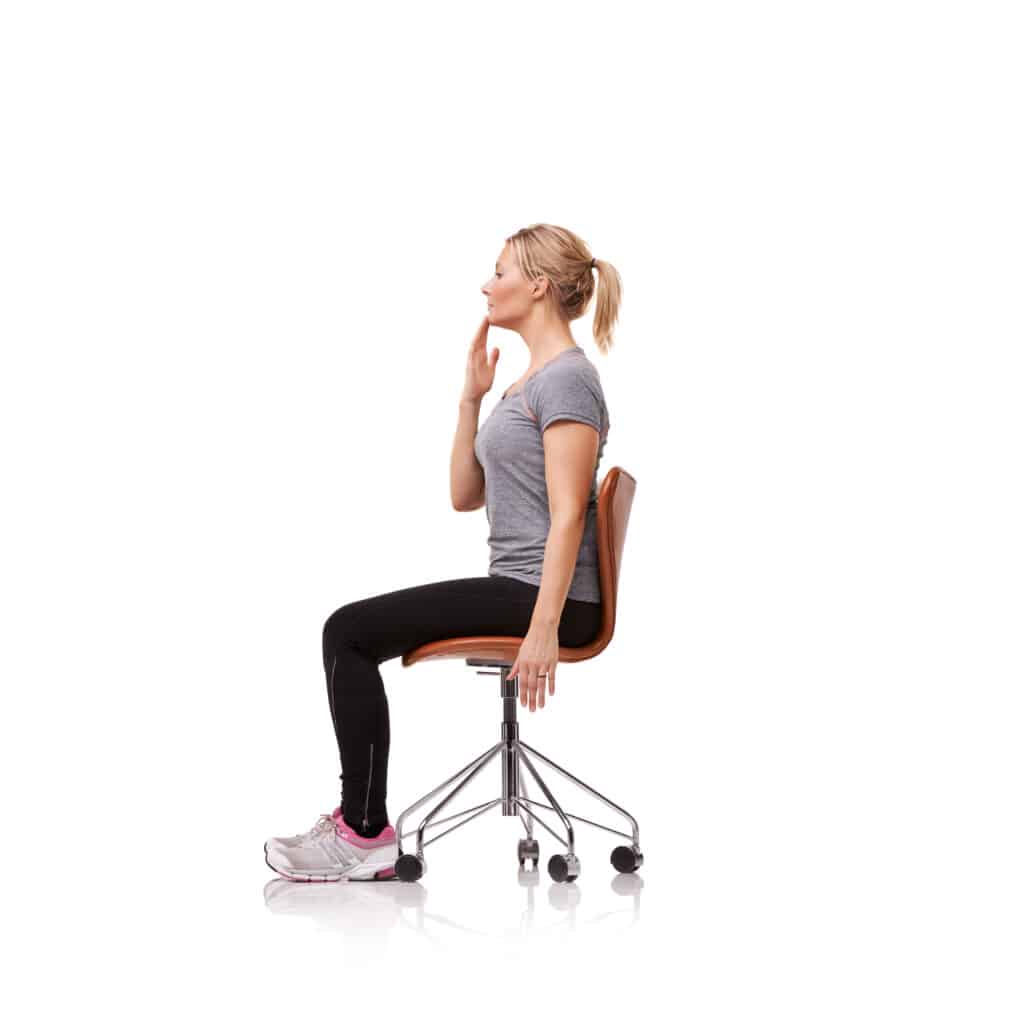
2) Upper Trapezius Stretch
Sit tall and hold the edge of your chair with your right hand. Tilt your left ear toward your left shoulder to feel a stretch on the right side of your neck.
Option: Rest your left hand lightly on the right side of your head for a gentle assist. Avoid pulling.
Hold 15 to 30 seconds, repeat 2 to 4 times each side.
3) Levator Scapulae Stretch
Turn your head about 45 degrees to the left, then tuck your chin toward your left armpit.
Place your left hand lightly on the back of your head to guide the stretch. You should feel it along the back and side of the neck.
Hold 15 to 30 seconds, repeat 2 to 4 times each side.
4) Doorway Chest Stretch
Tight chest muscles can pull the shoulders forward, which can increase neck strain.
Stand in a doorway with your forearms on the doorframe and elbows at shoulder height.
Step one foot forward and gently shift your weight until you feel a stretch across the chest and front of the shoulders.
Hold 15 to 30 seconds, repeat 2 to 3 times.
Back Stretches
These stretches focus on the upper and lower back, hips, and the muscles that support your spine.
1) Cat‑Cow Mobility
Start on hands and knees. Inhale as you gently let your belly lower and lift your chest. Exhale as you round your back and tuck your chin.
Move slowly through 8 to 12 repetitions.
2) Child’s Pose
From hands and knees, sit your hips back toward your heels and reach your arms forward.
Relax your shoulders and breathe into your sides and lower back.
Hold 20 to 30 seconds, repeat 2 to 3 times.
3) Single Knee‑to‑Chest
Lie on your back with knees bent. Bring one knee toward your chest, hands on the shin or behind the thigh.
Hold 15 to 30 seconds, then switch. Repeat 2 to 4 times each side.
4) Lower Trunk Rotations
Lie on your back with knees bent and feet flat. Gently let both knees fall to one side while keeping your shoulders on the floor.
Hold 10 to 20 seconds, then switch sides. Repeat 5 to 10 times total.
5) Prone Press‑Ups
The prone press up can help ease stiffness in the lower back for some people. If you feel more pain in your legs, numbness, tingling, or a change in how your legs or feet feel during the move, stop right away. Do not push through nerve symptoms, and talk with your clinician before trying again.
Lie on your stomach and prop up on your elbows, letting your low back relax.
Option: Press into your hands to raise your chest a bit higher if comfortable. Keep hips on the floor.
Hold 5 to 10 seconds, repeat 8 to 10 times.
6) Hamstring Stretch
Lie on your back. Loop a towel or strap around one foot and gently straighten the knee toward the ceiling until you feel a stretch in the back of the thigh.
Keep the other leg bent for comfort. Hold 15 to 30 seconds, switch sides, repeat 2 to 3 times.
7) Figure‑4 Hip Stretch
Lie on your back, cross your right ankle over your left knee. Lift the left leg and hold behind the thigh.
Feel the stretch in the right hip or buttock. Hold 15 to 30 seconds, switch sides, repeat 2 to 3 times.
How Often Should You Stretch?
Doing stretches regularly matters more than doing long, hard sessions. If you practice a little every day, you are more likely to keep your joints flexible and your back strong. Use the chart below to plan how often you stretch, how long to hold each stretch, and how many times to repeat.
Goal
Frequency
Hold Time
Repetitions
Ease stiffness
Daily or 5 days per week
15 to 30 seconds
2 to 4 per side
Posture support
Daily micro‑breaks
5 to 10 seconds for resets
Little and often throughout the day
Warm up
Before activity
Gentle, shorter holds
8 to 10 easy reps of mobility moves
Ergonomic Habits That Help
Set a reminder to stand and move for 1 to 2 minutes every 30 to 60 minutes.
Keep screens at eye level and your keyboard close so your shoulders stay relaxed.
Use a supportive chair, and place feet flat on the floor or on a small footrest.
Choose a pillow height that keeps your neck in a neutral position.
When To Contact Princeton Orthopaedic Associates
If your pain lasts more than a week or two despite trying home care, or if it wakes you at night or keeps you from daily tasks, you should seek an evaluation. Call if you notice numbness, tingling, weakness, or new changes in bowel or bladder control, as these need urgent attention.
Pain lasts more than 1 to 2 weeks despite home care.
Pain wakes you at night or limits daily tasks.
You notice numbness, tingling, or weakness in an arm or leg.
Pain began after a fall or accident.
There are changes in bowel or bladder control. This is urgent and needs immediate care.
Our team can diagnose the source of pain, tailor a stretching and strengthening plan, and coordinate physical therapy when needed.
Next Step
If your neck or back pain keeps returning, a personalized plan usually helps most. Schedule an exam with Princeton Orthopaedic Associates so we can identify what is driving your symptoms and guide you through the right exercises for long‑term relief.
This blog post is meant to be informative and should not act as a self-diagnosis tool. If you’d like to see one of our doctors, please contact us here.
What We Will Cover:
Understand your diagnosis and all treatment options.
Review the benefits, risks, and alternatives with your provider.
Follow all preoperative instructions regarding medications, diet, and fluid intake.
Be aware of what happens during the procedure and the safety checks involved.
Arrange for a ride home and prepare your space for a comfortable recovery.
Begin gentle movement and prescribed exercises on schedule.
Call your provider immediately if you experience any warning signs.
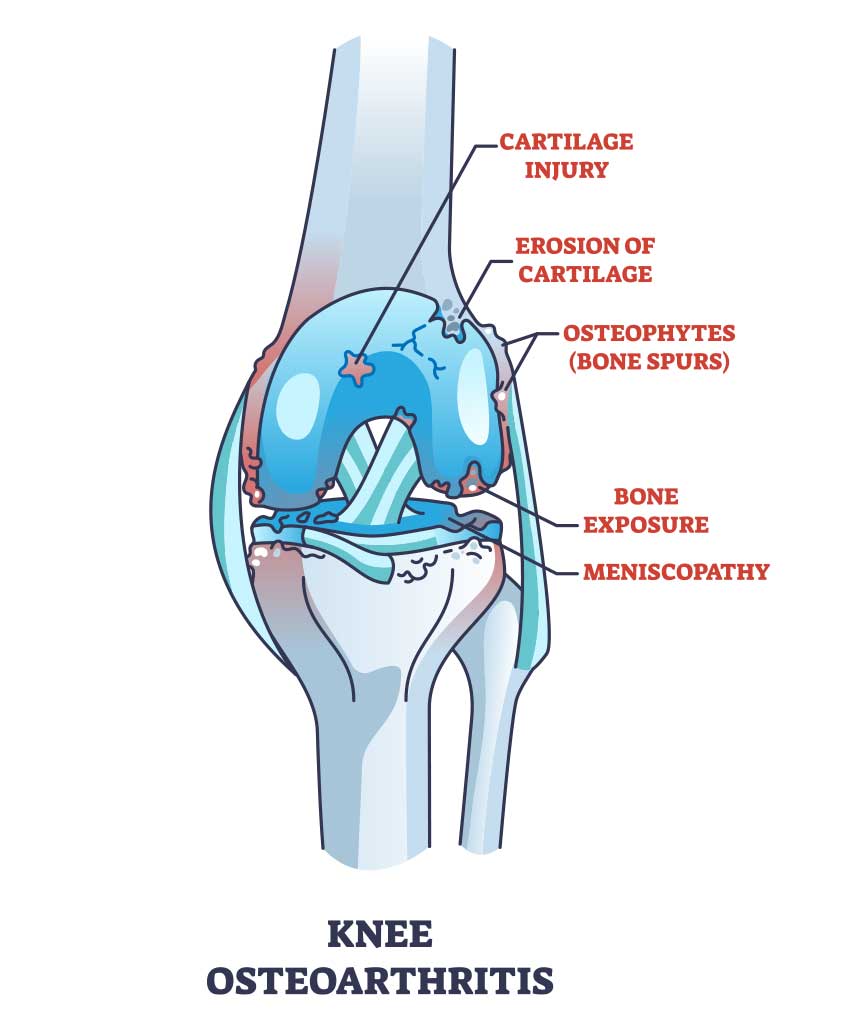
Understanding Your Knee Pain
Your knees support you through every step, bend, and climb. Over time, injuries or the natural wear of daily life can lead to pain, swelling, or a feeling of instability. If these symptoms begin to limit the activities you love, your orthopaedic surgeon may suggest knee arthroscopy, a minimally invasive procedure that uses small incisions to look inside the joint and treat the problem.
How the Knee Joint Works
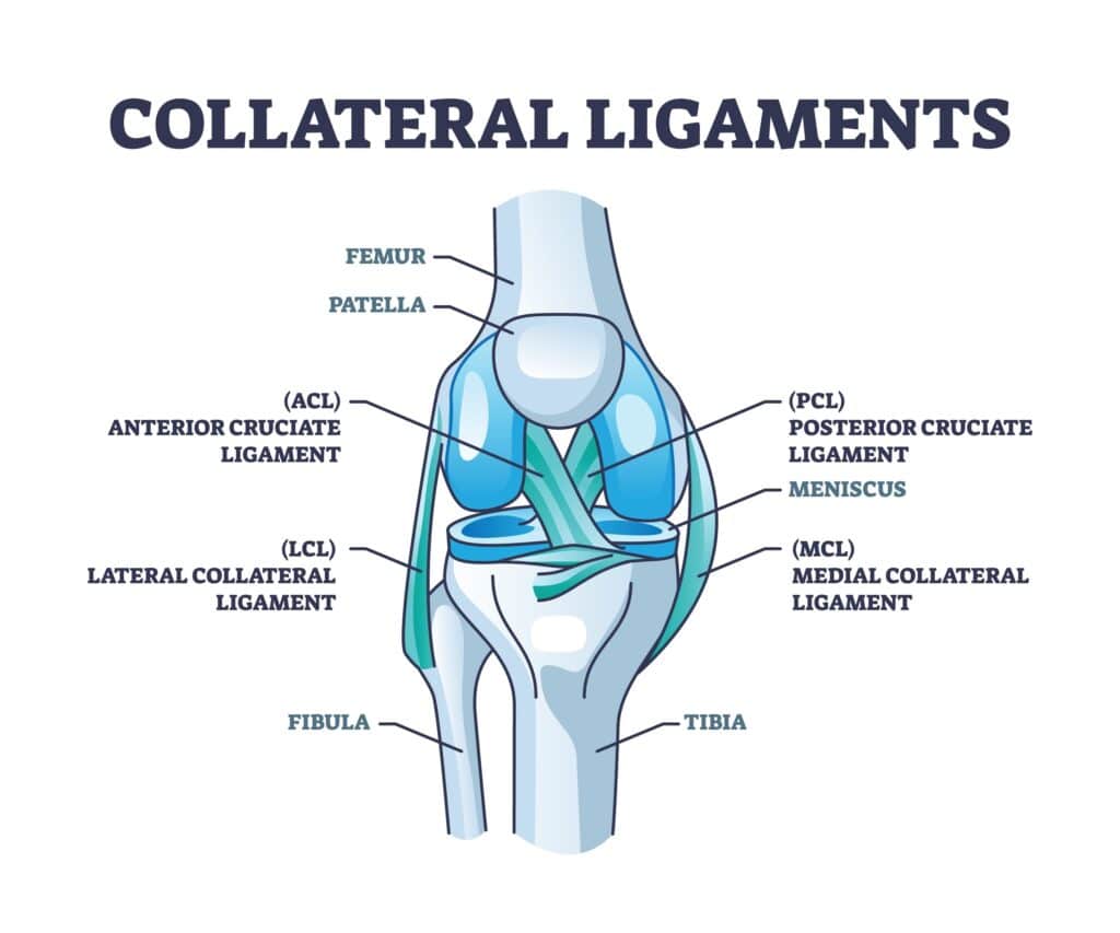
The knee is a complex hinge joint where your thigh bone (femur) and shin bone (tibia) meet. A C-shaped cushion of cartilage called the meniscus sits between these bones to absorb shock. The ends of the bones, along with the back of the kneecap (patella), are coated with a smooth, slick surface called articular cartilage, which allows the joint to glide comfortably. A network of muscles, tendons, and ligaments surrounds the joint to provide strength and stability.
Is Knee Arthroscopy Right for You?
To get an accurate diagnosis, your care team may order imaging tests like an X-ray or an MRI (magnetic resonance imaging). These tests can reveal issues in the soft tissues like ligaments and cartilage, as well as in the bones themselves. Arthroscopy can then be used to confirm what the images suggest and often allows your surgeon to treat the issue during the same procedure.
You and your provider will review the expected benefits and possible risks of arthroscopy. You will also discuss alternatives, which may include medications, physical therapy, activity changes, or wearing a brace. Be sure to ask every question on your mind so you feel informed and confident in your decision.
Your Knee Arthroscopy Procedure
What to Expect on Surgery Day
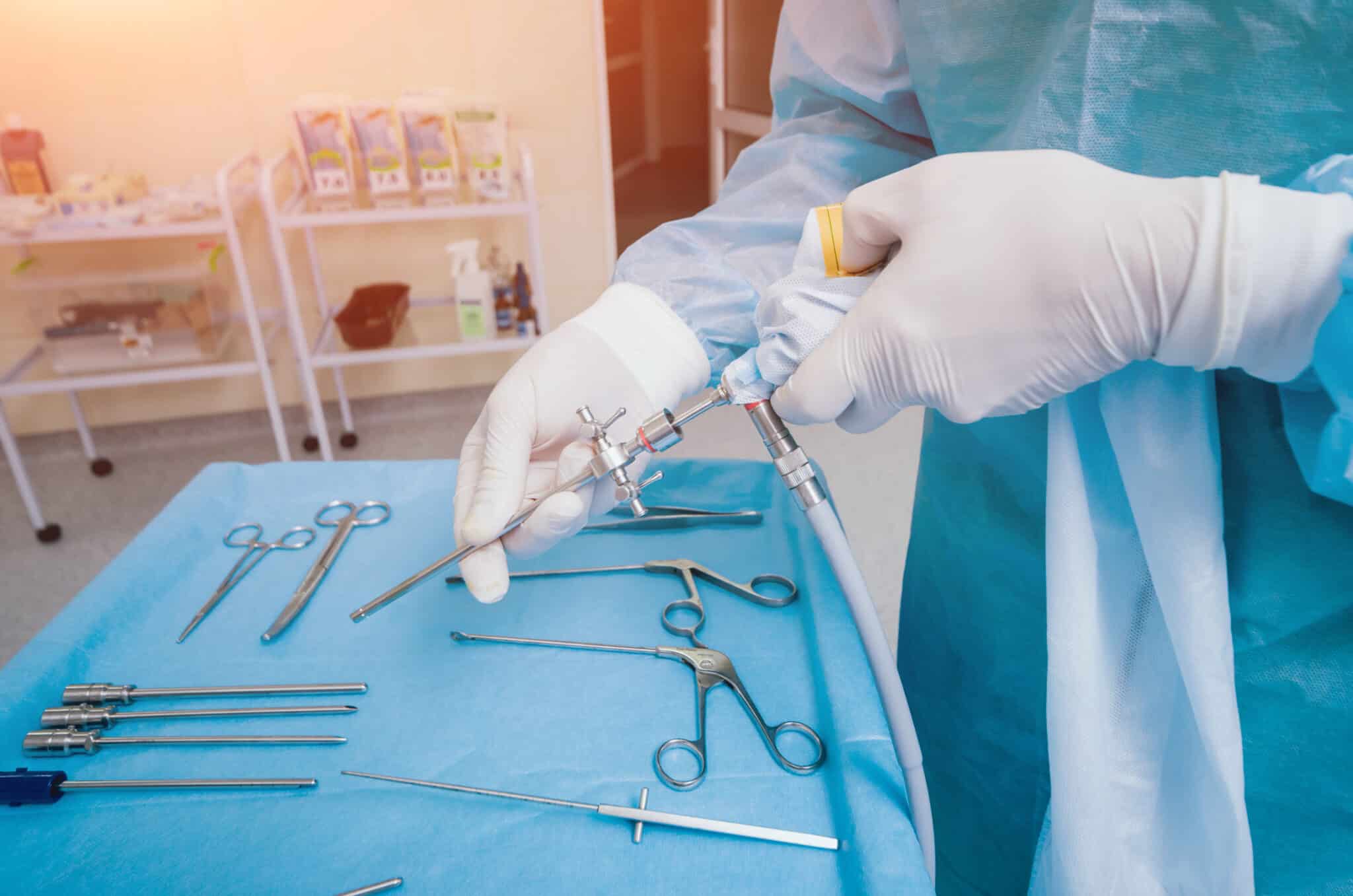
The Procedure: Arthroscopy allows the surgeon to see inside your knee using a thin, lighted camera called an arthroscope. The camera sends live images to a monitor, which guides the precise surgical tools inserted through separate small incisions. This approach allows the surgeon to diagnose and treat many knee problems through incisions that are typically less than an inch long.
Preparing: Before the procedure, inform your surgeon about all the medications and supplements you are taking. You may need to stop some of them ahead of time. You will also be instructed to stop eating and drinking for a set period to ensure your stomach is empty for anesthesia.
The Day of Surgery: On the day of surgery, your team will perform several routine safety checks, including marking the correct knee and confirming your identity and the planned procedure. Anesthesia will keep you comfortable and free from pain. The surgeon then makes two or three small incisions (called portals), places the scope through one, and inserts instruments through the others. Sterile fluid is used to gently expand the joint, improving visibility and helping the surgeon work accurately.
Risks and Possible Complications
Knee arthroscopy is a widely used and very safe procedure. However, like any surgery, it carries some risks. These may include:
Bleeding, infection, or blood clots
Stiffness or ongoing knee pain
Injury to blood vessels, nerves, or skin around the knee
Damage to cartilage, the meniscus, or ligaments
The need for additional surgery
Other specific risks as discussed by your surgeon
Common Conditions Treated with Arthroscopy
Arthroscopy can address a variety of common knee problems:
A Healthy Knee
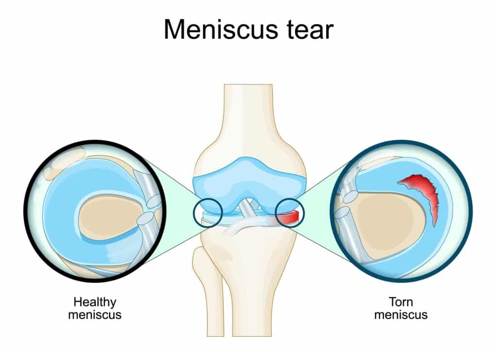
The Problem: Repeated squatting or a sudden twist can tear the meniscus. This may cause pain or swelling, and your knee may catch or lock when you move.
The Solution: Torn tissue on the inner portion of the meniscus is often trimmed away (a meniscectomy). Tears near the outer edge, which has a better blood supply, may be repaired with sutures.
Meniscus Tears
The Problem: Repeated squatting or a sudden twist can tear the meniscus. This may cause pain or swelling, and your knee may catch or lock when you move.
The Solution: Torn tissue on the inner portion of the meniscus is often trimmed away (a meniscectomy). Tears near the outer edge, which has a better blood supply, may be repaired with sutures.
ACL Tear
The Problem: A sudden pivot, cut, or awkward landing can tear the ACL. Patients often report a “pop,” rapid swelling within hours, and instability with pivoting or cutting.
The Solution: Low-demand or partial tears may be managed with structured rehab and bracing. Symptomatic complete tears, especially in patients returning to sports, are typically treated with arthroscopic ACL reconstruction using a tendon graft; primary ACL repair is reserved for select tears.
Cartilage Wear
The Problem: Articular cartilage can wear down, and loose pieces can float inside the joint. You may notice pain, stiffness, or a grinding sensation.
The Solution: The surgeon can remove loose fragments that irritate the joint to reduce catching and pain, and smooth down damaged cartilage surfaces.
Patella (Kneecap) Issues
The Problem: Articular cartilage can wear down, and loose pieces can float inside the joint. You may notice pain, stiffness, or a grinding sensation.
The Solution: The surgeon can remove loose fragments that irritate the joint to reduce catching and pain, and smooth down damaged cartilage surfaces.
Your Recovery at Home
Immediately After Surgery
When the procedure is finished, your small incisions will be closed and covered. In the recovery room, your knee will be bandaged, iced, and elevated to limit swelling. You will receive pain medication and be monitored by a nurse until it is safe to go home. Anesthesia and pain medicine can make you drowsy, so you must arrange for an adult to drive you home.
Caring for Your Knee
Follow your surgeon’s instructions carefully to ensure a smooth recovery. Key steps include:
Elevate your leg above the level of your heart as much as possible to reduce pain and swelling.
Ice your knee for 20-30 minutes several times a day for the first few days. Wrap the ice pack in a thin towel to protect your skin.
Keep incisions dry. Shower only when your provider says it is safe, and cover your leg with plastic to keep the bandages dry.
Manage weight-bearing. You may go home with crutches. Follow your weight-bearing directions carefully so your knee can heal properly.
Exercises for a Strong Recovery
Oftentimes, physical therapy is prescribed. But at home, you can work on many of the following.
Gentle movement is critical for healing. Your provider or physical therapist will guide you through a plan to restore motion and strength. Start these simple motions as soon as you are told it is safe to improve blood flow and help prevent blood clots.
Ankle Pumps: Point your foot down, then flex it up. Move your foot in circles several times throughout the day.
Quadriceps Sets: While lying down, tighten the muscles on the front of your thigh and press the back of your knee toward the surface. Hold for 5 to 10 seconds, then relax. Repeat as directed.
Straight Leg Raises: Lying down, keep your knee straight and lift your leg 8 to 12 inches off the surface. Hold for 5 seconds, then lower slowly. Repeat as directed.
When to Call Your Provider
Contact your provider’s office right away if you notice any of the following:
Fever of 100.4° F (38° C) or higher
Pain that does not improve with medication and rest
Swelling that does not improve with elevation and icing
Increased redness, warmth, or drainage from the incision sites
Bleeding that soaks through your bandages
New or worsening numbness in your leg or foot
Severe nausea or vomiting
Returning to Your Activities
Recovery time varies based on your specific procedure, the condition of your knee, and your overall health. Many people with desk jobs can return to work in about one week. Jobs that require prolonged standing or heavy activity may require more time off. With consistent effort in your rehabilitation, most people can return to their normal active lifestyle within one to two months.
Knee arthroscopy is a powerful tool for diagnosing and treating the cause of your knee pain. By preparing as instructed and following your post-operative plan closely, you give yourself the best chance of returning to your activities with less pain and improved function.
This blog post is meant to be informative and should not act as a self-diagnosis tool. If you’d like to see one of our doctors, please contact us here.