You know the pain well. As you rise from bed every morning, you place your feet on the floor, only to be greeted with a sharp, stabbing sensation in your heel. It's like a jolt that reminds you that the pain isn't gone yet. After walking around for a few minutes, the discomfort may ease. Still, it always comes back when you've been on your feet too long, especially after resting or sleeping. This is a daily struggle for those living with plantar fasciitis – a condition that affects your ability to walk comfortably and can take a toll on your overall quality of life.
What is Plantar Fasciitis?
Plantar fasciitis occurs when a thick connective tissue running from your heel to your toes along the bottom of your foot (plantar fascia) becomes irritated and inflamed. The plantar fascia supports the foot's arch and is essential for proper foot movement and support while walking. Excessive strain or overstretching can cause tiny tears in the fascia, triggering inflammation and discomfort, particularly in the heel.
While plantar fasciitis is frequently seen in physically active individuals, it can affect anyone. Symptoms tend to worsen in the morning when the tissue tightens during sleep, causing significant discomfort with the day's first steps.
Who Gets Plantar Fasciitis?
If you’ve ever felt a sharp, stabbing pain in your heel when you step out of bed in the morning, you may be dealing with plantar fasciitis—one of the most common causes of heel pain. This condition affects millions of people each year, but it’s not random. Certain groups are more likely to develop it based on lifestyle, footwear choices, and physical activity.
1. Runners and Athletes
People who run regularly or participate in high-impact sports are among the most common groups to experience plantar fasciitis symptoms. Repetitive motion, especially without proper stretching or supportive footwear, can cause microtears in the plantar fascia. Increasing mileage too quickly, running on hard surfaces, or using worn-out shoes can all contribute to heel pain in runners.
2. People Who Stand All Day
Working a job that keeps you on your feet for hours at a time can put you more at risk for developing plantar fasciitis. Teachers, nurses, warehouse workers, and retail staff often experience chronic foot and heel pain due to the constant pressure on their feet—especially if they’re standing on hard surfaces like concrete or not wearing cushioned shoes.
3. Those with Excess Body Weight
Carrying extra weight puts more pressure on your feet and heels, making it harder for your plantar fascia to do its job. This is especially true during long periods of walking or standing. Losing even a modest amount of weight can often reduce pain and prevent further damage.
4. People with Foot Structure or Gait Issues
Your natural foot shape or the way you walk could be behind your chronic heel pain. People with flat feet, high arches, or those who overpronate (roll their feet inward too much) often experience more strain on the plantar fascia, leading to irritation and inflammation. Tight calf muscles or a limited range of motion in your Achilles tendon can also increase your risk.
5. Adults Between Ages 40–60
Unfortunately, age is another common factor. Middle-aged adults are more likely to develop plantar fasciitis due to natural wear and tear. The connective tissue in our bodies loses elasticity as we age, making us more prone to injury from overuse or poor support.
6. People Who Wear Unsupportive Shoes
Let’s talk about your footwear! Flip-flops, ballet flats, high heels, and worn-out sneakers all share the same issue, they offer little to no arch support. Wearing these types of shoes regularly can increase your risk of developing plantar fasciitis, especially if you're walking long distances or spending hours on your feet.
Why you can trust us:
We have multiple highly specialized, board-certified, fellowship-trained orthopaedic surgeons.
We know that we serve people - actual humans - not random orthopaedic conditions. That drives us to compassionate care.
The world of orthpaedics is constantly evolving. Our orthopaedic surgeons are constantly evaluating new techniques, tools and methods to serve our community even better.
We provide outcome-focused treatment. We work with our patients to achieve their goals, all while developing custom treatment plans that fit our patient's lives.
The symptoms of plantar fasciitis can vary in intensity but generally include:
Heel pain – The most common symptom is a sharp pain, often felt in the center or at the bottom of the heel. It can feel like a stabbing sensation, making walking difficult, especially after resting.
Pain after rest – The pain is typically more severe after long periods of sitting or lying down, such as when you first get out of bed or after long periods of inactivity.
Swelling: Mild swelling may occur at the bottom of the heel, especially after standing for extended periods.
Pain with prolonged standing or walking may decrease as you walk. Still, after prolonged periods of standing or after physical activity, the pain can return and become more intense.
Stiffness – After sitting or lying down for a long time, the bottom of the foot can feel stiff, making it difficult to take the first few steps of the day.
Causes of Plantar Fasciitis
Several contributing factors can lead to plantar fasciitis. Understanding these causes of plantar fasciitis can help prevent the condition or reduce the risk of recurrence:
Overuse and repetitive stress
Plantar fasciitis is often caused by repetitive activity that stresses the plantar fascia. High-impact activities like running or jumping can overstrain this tissue, especially if done incorrectly or without proper footwear.
Improper footwear
Shoes that lack adequate arch support, cushioning or are worn-out can exacerbate heel pressure. High heels, flip-flops, or shoes with flat soles may contribute to this strain.
Foot structure abnormalities
People with flat feet, high arches, or abnormal gait patterns may place excessive stress on the plantar fascia. The added strain causes the tissue to become overstretched, leading to inflammation and pain.
Obesity or excess weight
Extra weight puts additional pressure on the feet, making them more susceptible to injury and strain. Over time, this additional weight can cause microtears in the plantar fascia, leading to plantar fasciitis.
Tight calf muscles or Achilles
Having tightness in the calves or Achilles tendon can affect how pressure distributes across your feet. This adds stress to the plantar fascia, increasing the likelihood of inflammation and discomfort.
Aging
As you age, the plantar fascia loses some elasticity and shock-absorbing properties. This makes the tissue more prone to tears and inflammation, leading to plantar fasciitis, especially in people over 40.
When to See an Orthopedic Specialist For Plantar Fasciitis
You can usually manage plantar fasciitis with at-home treatments. Still, sometimes, certain situations warrant a visit to an orthopedic specialist.
Signs to see an orthopaedic specialist for plantar fasciitis:
Persistent pain – If your plantar fasciitis pain lasts more than a few weeks despite your self-care efforts, it's time to seek professional help. Chronic pain could mean the inflammation has not subsided and may require more aggressive treatment.
Difficulty walking – If the pain interferes with your ability to walk or complete daily tasks, seeking care from an orthopedic specialist is essential.
No improvement with over-the-counter treatments – If over-the-counter pain relievers or home remedies like ice and stretching haven't brought any relief, you might need more specialized care to resolve the issue.
Swelling and bruising – If you experience swelling, bruising, or warmth around the heel, it could signal more than just plantar fasciitis. An orthopedic specialist will perform diagnostic tests to ensure no other underlying injury.
Nerve damage symptoms – If you experience numbness, tingling, or weakness in your foot, this could be a sign of nerve involvement, which requires immediate attention.
How to Treat Plantar Fasciitis at Home
Here are several strategies you can use at home to help relieve the pain and inflammation associated with plantar fasciitis:
Rest and avoid high-impact activities
Giving your foot time to heal is essential. Avoid activities that involve running, jumping, or standing for long periods, as these can further irritate the plantar fascia.
Ice therapy
Applying an ice pack to your heel for 15-20 minutes several times throughout the day can help reduce inflammation and pain.
OTC anti-inflammatory medications
Medications like ibuprofen or naproxen can reduce inflammation and relieve pain. Be sure to follow the recommended dosage and consult a doctor if needed.
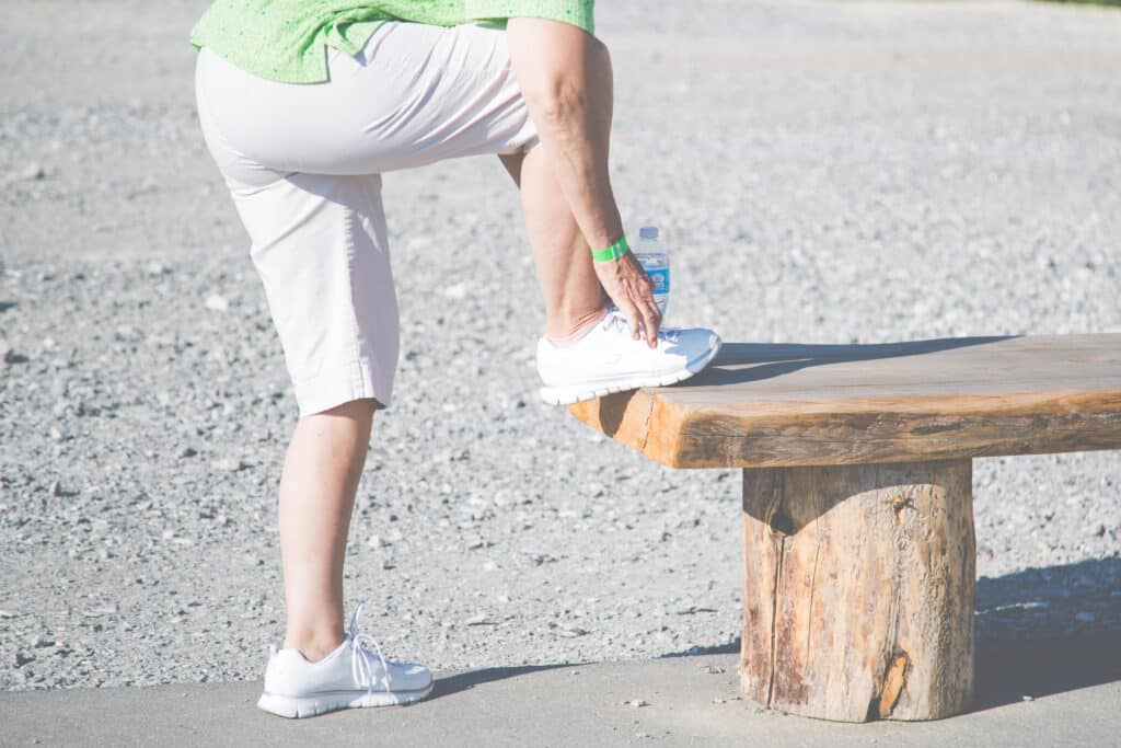
Stretching exercises
Stretching your calf muscles, Achilles tendon, and the plantar fascia can help alleviate your pain and prevent stiffness. Simple exercises like towel stretches or calf stretches can improve your flexibility and reduce the strain on your foot.
Foot supports
Wearing supportive shoes with proper arch support and cushioning can reduce pressure on your plantar fascia. Orthotic insoles may also help distribute pressure more evenly across your foot.
Night splints
Wearing night splints can help maintain a light stretch of the plantar fascia while you sleep. This helps prevent the fascia from tightening during the night and reduces morning pain.
How to Prevent Plantar Fasciitis
Preventing plantar fasciitis is possible but it involves making adjustments to your daily habits and lifestyle.
How to prevent plantar fasciitis:
Wear proper footwear – Select footwear that provides the right arch support, cushioning, and stability for your heels. Avoid walking barefoot on hard surfaces and limit prolonged use of high heels to reduce strain on your feet.
Regular Stretching – Regularly stretch your calves, Achilles tendons, and feet to improve flexibility and reduce the risk of strain on the plantar fascia.
Maintain a healthy weight – Keeping your weight within a healthy range will help to reduce the strain on your feet, decreasing your chances of developing plantar fasciitis.
Take breaksto rest – If you stand or walk for long periods, take breaks to rest your feet and alleviate pressure. If you can't take breaks, alternating between sitting and standing can also help prevent overuse.
Strengthen your feet and lower legs – Exercises that strengthen your calf muscles, foot muscles, and ankle stabilizers can help prevent plantar fasciitis by improving foot mechanics and reducing excessive strain.
Exercises to Help Release and Heal Plantar Fasciitis
Here are some targeted exercises to help with plantar fasciitis:
Towel Stretch – Sitting on the floor with your legs stretched out in front of you, wrap a towel around the ball of your foot. Gently pull your foot toward you, feeling a stretch along the bottom of your foot and the back of your leg.
Calf Stretch – Stand arm's length facing a wall and step back with one leg. Keep your back leg straight and your heel planted on the ground. Place your hands on the wall at the height of your shoulders and lean forward, stretching your calf and Achilles tendon of your back leg.
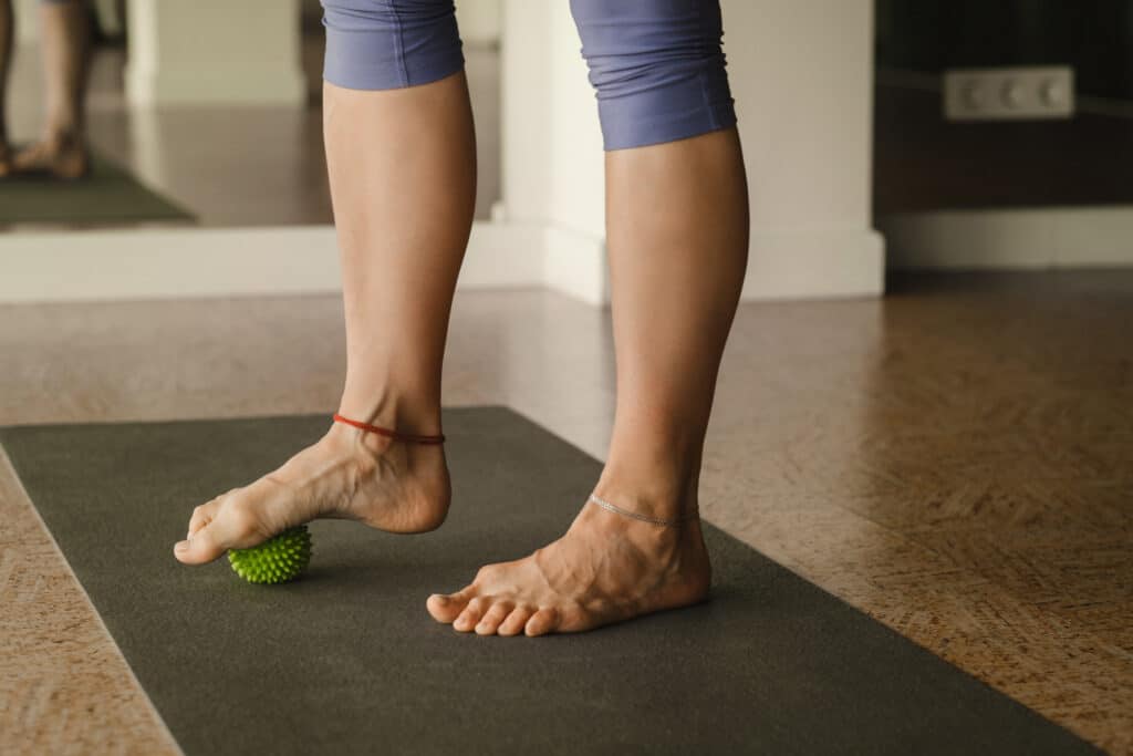
Foot Roll – To help massage the plantar fascia and relieve built-up tension, you can use a tennis ball or similar ball under the arch of your foot. Once you place it, slowly roll it back and forth.
Toe Stretch – While sitting with your legs extended, reach for your toes and gently pull them toward your body. This stretch helps loosen the plantar fascia and should be held for 15-30 seconds before repeating.
Heel Raises – Stand with the balls of your feet on the edge of a step. Slowly lift yourself onto your toes, then lower your heels past the level of the step to stretch your calves and the bottom of your feet.
Whether you're a runner, a retail worker, or someone who loves their flip-flops a little too much, plantar fasciitis can sneak up on you. Knowing your risk factors is half the battle. With the right footwear, regular stretching, and paying attention to heel pain symptoms, you can take steps (literally!) to protect your feet.
Plantar fasciitis doesn't have to be a permanent issue. You can effectively manage and treat this painful condition with proper care, early intervention, and lifestyle adjustments. If your at-home treatments aren't bringing you relief or if your symptoms are worsening, Princeton Orthopaedic Associates is here to help. Our team of specialists can provide targeted therapies, advanced treatments, and personalized care to help you get back on your feet and live pain-free.
This blog post is meant to be informative and should not act as a self-diagnosis tool. If you’d like to see one of our doctors, please contact us here.
Morton's Neuroma: What It Is and How To Manage It
What Is Morton's Neuroma?
If you've ever felt like you're walking on a pebble, experienced burning pain in the ball of your foot, or a stabbing pain between your toes, you may be dealing with a condition known as Morton's neuroma. Morton's neuroma happens when the tissue around one of the nerves leading to one of your toes becomes compressed or irritated, leading to thickening and inflammation. This thickening causes you pain and discomfort, especially when walking. The nerve between the third and fourth toes is often affected, though it can happen elsewhere in the foot.
Symptoms of Morton's Neuroma
Morton's neuroma symptoms can sneak up on you. The condition can start with subtle discomfort and gradually progresses if left untreated.
Here are the hallmark symptoms of Morton's neuroma to look out for:
Relief when removing shoes, staying off your feet, or massaging the foot.
A feeling of standing on a pebble or bunched-up sock.
A sharp, burning pain in the ball of your foot or between your toes.
Tingling or numbness in your toes around the affected nerve.
Pain that worsens when wearing tight shoes or high heels.
10 Signs You May Have Morton's Neuroma
Early recognition of the signs of Morton's Neuroma can help you prevent worsening pain and complications. Here are 10 key indicators that you might be dealing with Morton's neuroma:
Having persistent pain in the ball of your foot.
Experiencing tingling or numbness in your toes.
Feeling a sensation of clicking or popping when pressing on the ball of your foot or when walking.
Experiencing discomfort while walking or running.
Having pain that radiates into your toes.
Having symptoms that improve when barefoot but worsen in shoes.
Experiencing burning or stabbing pain localized between your toes.
Seeing visible swelling in the ball of the foot (less common).
Having difficulty wearing tight or narrow footwear.
Experiencing symptoms that are gradually worsening over time.
Learn more about POA's Podiatrists:
Why you can trust us:
We have multiple highly specialized, board-certified, fellowship-trained orthopaedic surgeons.
We know that we serve people - actual humans - not random orthopaedic conditions. That drives us to compassionate care.
The world of orthpaedics is constantly evolving. Our orthopaedic surgeons are constantly evaluating new techniques, tools and methods to serve our community even better.
We provide outcome-focused treatment. We work with our patients to achieve their goals, all while developing custom treatment plans that fit our patient's lives.
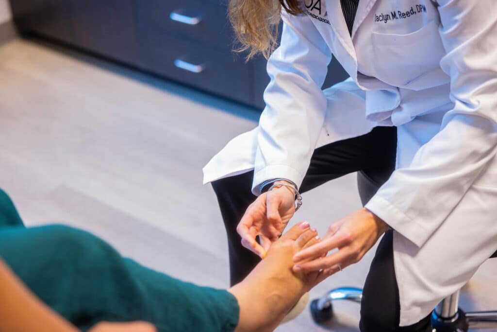
Getting an accurate diagnosis is the first step toward relief. During a medical evaluation, your doctor will consider your symptoms and may perform specific tests. These evaluations often include:
Pressing on the ball of your foot to check for pain or clicking.
Review your medical history and symptoms.
Imaging tests like an X-ray, ultrasound, or MRI may be recommended to rule out other conditions.
At POA, we have a team of doctors who specialize in foot issues and injuries. These podiatrists see patients with foot conditions all day, every day. That makes them foot experts, and that's the quality of world-class care you will find at Princeton Orthopaedic Associates.
What Else Could be Causing Pain Between My Toes
Several conditions can mimic the symptoms of Morton's neuroma but are distinctly different. These possible conditions include:
Metatarsalgia: A general term for pain and inflammation in the ball of the foot, often caused by overuse or improper footwear.
Stress Fractures: Tiny cracks in the bones of the foot, typically due to repetitive stress or overuse, causing localized pain.
Capsulitis: Inflammation of the ligaments surrounding the joints at the base of the toes, particularly the second toe, causing pain similar to Morton's neuroma.
Tarsal Tunnel Syndrome: A condition involving compression of the posterior tibial nerve as it passes through the tarsal tunnel near the ankle, leading to pain and tingling in the foot.
Plantar Plate Tear: A tear or weakening of the ligament beneath the toes, usually the second toe, causing pain and instability in the ball of the foot.
Bursitis: Inflammation of the bursae (fluid-filled sacs) in the foot, which can cause pain and discomfort in areas similar to Morton's neuroma.
Ganglion Cysts: Fluid-filled lumps that can form on the nerves or tendons in the foot, sometimes causing pain or discomfort.
Each of these conditions has unique characteristics and requires different treatment approaches. If you're having foot pain, we recommend scheduling a time with one of our specialists. Having the correct diagnosis will set you on the path to the quickest recovery.
Morton's Neuroma Treatment
If you're diagnosed with Morton's neuroma, the good news is that there are several treatment options available. From conservative approaches for you to address your Morton's neuroma at home to more involved procedures, here are some common ways to address the condition:
Footwear Changes: Switch to shoes with a wider toe box and avoid high heels.
Orthotic Inserts: Custom or over-the-counter arch supports can relieve pressure on the nerve.
Padding: Metatarsal pads can reduce stress on the ball of your foot.
Medications: Nonsteroidal anti-inflammatory drugs (NSAIDs) help reduce pain and inflammation.
Corticosteroid Injections: These can provide significant relief by reducing swelling around the nerve.
Physical Therapy: Specific stretches and exercises can improve foot function.
Radiofrequency Ablation: A minimally invasive procedure to block pain signals from the affected nerve.
Surgery: In severe cases, surgery may be recommended to remove the neuroma.
Can I Cure My Morton's Neuroma?
Morton's neuroma can feel overwhelming, but many people have successfully managed or eliminated their symptoms with consistent effort and self-care measures. Taking proactive steps at home can help you manage Morton's neuroma and prevent symptoms from worsening. Consider these simple yet effective self-care strategies for addressing your Morton's neuroma.
Switching Footwear: Wear shoes with low heels and plenty of cushioning. Investing in wide, supportive shoes with good arch support is key!
Using Orthotics: Custom inserts reduced pressure on the ball of the foot.
Stretching: Regular exercises to stretch the toes and foot muscles.
Rest: Rest your feet and avoid activities that worsen your pain.
Ice: Use ice packs to reduce swelling; apply ice packs for 15-20 minutes multiple times a day.
Professional Treatment: Consult with a foot specialist to create a tailored plan for finding relief during flare-ups.
Morton's Neuroma Exercises
Regular exercises can help alleviate your Morton's neuroma symptoms and improve your overall foot strength and health. The following exercises are easy to perform, and you can do them at home. Some of the most effective include:
Toe Spreading: Place a rubber band around your toes and spread them apart.
Ball Rolls: Roll a tennis ball under the arch of your foot. You can also freeze a water bottle and use that to roll under the arch of your foot.
Toe Stretches: Use your hands to gently pull your toes backward to stretch the ball of your foot.
Calf Stretches: Stretching your calves reduces tension on the foot.
Best Shoes for Morton's Neuroma
Shoes can absolutely make or break your foot comfort and health, especially when dealing with Morton's neuroma. Choosing the right footwear can drastically reduce discomfort.
Here's what to look for in the best shoes for people with Morton's neuroma:
Wide Toe Box: Avoid narrow or pointy shoes.
Cushioned Soles: Look for soft, supportive soles that absorb shock.
Low Heels: Flat or low-heeled shoes reduce pressure on the ball of the foot.
Adjustable Straps or Laces: Ensure a snug but comfortable fit.
Arch Supports and Inserts for Morton's Neuroma
Orthotics can provide essential support for your feet and they can also help to relieve pressure on the affected nerve causing your Morton's neuroma symptoms. There are different types of orthotics, each offering unique benefits:
Custom Orthotics: Tailored to your foot shape for maximum comfort and support of the entire foot.
Over-the-Counter Inserts: Although OTC orthotics are not custom to your specific foot, many more affordable options can provide adequate arch support for mild cases.
Metatarsal Pads: Specifically designed to alleviate stress on the ball of the foot.
If you suspect you have Morton’s neuroma due to persistent foot pain, it’s important to see a podiatrist for evaluation and treatment. If your symptoms worsen over time, interfere with daily activities, or do not improve with home remedies such as rest, ice, proper footwear, or over-the-counter pain relief, a podiatrist can provide a professional diagnosis. They may use imaging tests, recommend custom orthotics, or suggest more advanced treatments like corticosteroid injections or minimally invasive procedures if necessary. Early intervention can prevent the condition from worsening and improve your overall foot health.
When to Consider Morton's Neuroma Surgery
In some cases, conservative treatments aren't enough, and surgery becomes the best option. If your pain is severe or persistent, it may be time to discuss surgical solutions with your doctor.
Surgery may be necessary if:
Non-surgical treatments fail to provide relief.
The neuroma significantly interferes with daily activities.
Pain continues to worsen despite treatment.
Surgical options include removing the neuroma or releasing the surrounding ligament to reduce pressure on the nerve. Recovery can typically involve a few weeks of limited activity; however, each person is unique, so your treatment and recovery will be specific to you. Every person's experience with Morton's neuroma is specific to them, and surgery may not be necessary for you. Seeking professional advice from one of POA's podiatrists will help you get the best treatment possible.
No More Morton's Neuroma Foot Pain!
Your Morton's neuroma pain doesn't have to keep you from doing the things you need to in your daily life. It is possible for you to manage or even resolve your symptoms with proper care, which can include the right footwear, exercises, support, and professional guidance. If you are dealing with recurrent or persistent pain in the ball of your foot and think you have Morton's neuroma, consult with one of our podiatrists or explore your options so you can get one step closer to being pain-free.
Need Care Now? POA Has Six Urgent Care Facilities
Monroe
11 Centre Drive Monroe Twp., NJ 08831
Plainsboro
5 Plainsboro Road, Suite 100 Plainsboro, NJ 08536
Robbinsville
1 Union Street Suite 305 Robbinsville, NJ 08691
Princeton
325 Princeton Avenue Princeton, NJ 08540
Hillsborough
315 US Highway 206 Hillsborough Township, NJ 08844
This blog post is meant to be informative and should not act as a self-diagnosis tool. If you’d like to see one of our doctors, please contact us here.
Experiencing a stress fracture often means dealing with persistent pain that worsens with activity and eases with rest. You might notice swelling and tenderness at the injury site, making everyday tasks increasingly uncomfortable. Simple actions like walking or standing for long periods become challenging, and you might be reluctant to put weight on the affected limb. Missteps or sudden movements can cause sharp, shooting pain, keeping you constantly aware of the injury.
What is a Stress Fracture?
A stress fracture is a tiny crack in a bone that is caused by repetitive force, often from overuse. Unlike a sudden break from a single injury, stress fractures develop gradually over time, making them a common issue for athletes and individuals engaged in repetitive activities.
Where Does a Stress Fracture Occur?
Stress fractures can occur in any bone subjected to repetitive stress, but they are most commonly found in:
Metatarsals: The long bones in the foot, especially the second and third metatarsals.
Tibia: The shinbone, particularly in runners.
Femur: The thigh bone, especially in its neck (near the hip).
Fibula: The smaller bone in your lower leg.
Navicular: A bone on the top of the midfoot.
Pelvis: The hip area, particularly in female athletes.
Lumbar Spine: Lower back, particularly in gymnasts and dancers.
How Does a Stress Fracture Happen?
Stress fractures are primarily caused by overuse and repetitive activities. Factors contributing to stress fractures include:
Sudden Increase in Activity:
A rapid uptick in the frequency, duration, or intensity of your physical activity without adequate preparation.
Improper Footwear:
If you are wearing shoes that do not provide your foot with proper support or if they are worn out.
Poor Technique:
Incorrect form during physical activities can place undue stress on bones.
Low Bone Density:
Conditions like osteoporosis can make bones more susceptible to fractures.
Insufficient Rest:
Not allowing the body enough time to recover between activities.
What Does a Stress Fracture Feel Like?
Stress fractures typically start with a dull, localized pain that gradually worsens with activity and improves with rest. Key symptoms include:
Pain: Pain during weight-bearing activities that subsides with rest.
Swelling: Mild to moderate swelling at the fracture site.
Tenderness: Point tenderness directly over the fracture site.
Bruising: Occasionally, mild bruising may be present.
Why You Shouldn't Ignore a Stress Fracture
Ignoring a stress fracture can lead to serious complications and prolonged healing:
Complete Fracture: The tiny crack can develop into a complete break if the bone continues to be stressed.
Delayed Healing: Without proper rest and treatment, the fracture may not heal properly, leading to chronic pain and potential deformities.
Secondary Issues: Compensating for pain can lead to other injuries, such as muscle strains and joint problems.
Daily Life With a Stress Fracture
Living with a stress fracture can be challenging. Activities you once took for granted, like walking or climbing stairs, can become painful. Simple tasks can aggravate the pain, forcing you to modify your routine significantly. The pain and limitations can also affect your mental health, causing frustration and anxiety about your recovery and future activity levels.
Imagine you are an avid runner, and suddenly, every step feels like a sharp pain in your shin. You might find yourself unable to participate in your daily jog, impacting your fitness routine and social interactions. Even walking your dog or going to the grocery store can become daunting tasks. The frustration of having to sit out from activities you love can take a toll on your mental well-being, adding to the physical discomfort.
Need Care Now? POA Has Six Urgent Care Facilities
Monroe
11 Centre Drive Monroe Twp., NJ 08831
Plainsboro
5 Plainsboro Road, Suite 100 Plainsboro, NJ 08536
Robbinsville
1 Union Street Suite 305 Robbinsville, NJ 08691
Princeton
325 Princeton Avenue Princeton, NJ 08540
Hillsborough
315 US Highway 206 Hillsborough Township, NJ 08844
Seeking care from a specialist early on can help with proper diagnosis and a faster recovery. Here’s what to expect:
Diagnosis
Physical Examination: Your orthopaedic specialist will check for tenderness and swelling in the affected area.
Imaging Tests: X-rays, MRIs, or bone scans may be used to confirm the diagnosis of a stress fracture and assess the severity.
Common Stress Fracture Treatment Options
Rest
Rest is usually the primary treatment for stress fractures. This means avoiding activities that put stress on the affected bone. For a runner, this might mean taking a break from running and engaging in low-impact activities like swimming or cycling until the bone heals.
Protective Footwear or Bracing
Special shoes or braces can help offload stress from the fracture site. For example, a walking boot can protect a stress fracture in the foot, allowing you to move around without putting pressure on the injured area.
Physical Therapy
Physical therapy with exercises and stretches designed to improve your strength and flexibility in the affected area can help you heal. Our physical therapists can create a personalized program to help you recover safely. They might include exercises that gradually increase in intensity, ensuring the bone heals properly without being overstressed.
Once the pain has subsided and the bone shows signs of healing, you can gradually reintroduce activities. Start with low-impact exercises and slowly build up to your previous activity level. This helps prevent re-injury and ensures a smooth transition back to your regular routine.
Surgery
For a severe stress fracture, surgical intervention may be necessary to stabilize the fracture. This might involve inserting metal pins or screws that will hold the bone together as it heals. Surgery is typically considered when conservative treatments fail or when the stress fracture is in a high-risk area that is less likely to heal on its own.
Preventing stress fractures involves several strategies:
Gradual Increase in Activity: Increase your activity level gradually to avoid overloading your bones.
Proper Footwear: Invest in shoes that provide adequate support and replace them regularly.
Cross-Training: Engaging in a variety of workouts and physical activities will help you avoid repetitive stress on the same bones and can reduce the chances of another stress fracture.
Strength Training: Incorporating strength training exercises can help you to improve muscle support around the bones.
Adequate Nutrition: Ensure you get enough calcium and vitamin D to support bone health.
Stress fractures are a common but serious injury resulting from repetitive stress on bones. They require timely diagnosis and treatment to prevent further complications. Ignoring a stress fracture can lead to complete breaks, delayed healing, and secondary issues. If you suspect you have a stress fracture, you should seek medical advice promptly to ensure a full recovery and a return to your normal activities. By understanding the symptomsof stress fractures, causes, and treatment options, you can take proactive steps to prevent stress fractures in the future.
Living with a stress fracture can significantly impact your daily life, making it essential to address the injury promptly. Proper treatment and prevention strategies can help you recover fully and get back to your active lifestyle. Contact us today.
Understanding Bone Spurs
Have you ever experienced a sharp pain in your foot when taking your first steps in the morning or a nagging ache in your shoulder that won't go away? You might be dealing with a foot or shoulder bone spur. These small, bony growths can develop in various parts of the body, causing discomfort and limiting your mobility.
What is a Bone Spur?
A bone spur, also known as osteophyte, is a bony projection that forms along the edges of bones. These growths typically develop where bones meet each other in the joints. While bone spurs are not necessarily painful, they can cause problems when they rub against nearby nerves or tissues.
What Causes Bone Spurs?
Bone spurs often develop in response to pressure, rubbing, or stress on a bone over time.
Common causes include:
Osteoarthritis
Osteoarthritis is a degenerative joint disease and one of the most common causes of bone spurs. As the protective cartilage between bones wears down over time, the body may respond by forming extra bone around the affected joint edges, resulting in bone spurs.
Repetitive Stress or Overuse
Activities that involve repetitive motions or stress on specific joints, such as regularly lifting heavy objects, running, or jumping, can lead to the formation of bone spurs. Over time, the constant pressure on the bones can cause them to develop extra bony growths.
Age-related Wear and Tear
As people age, the cartilage in their joints naturally begins to deteriorate. This can result in increased bone friction, leading to bone spurs, especially in weight-bearing joints like the spine, knees, or hips.
Joint Diseases
Inflammatory joint conditions such as rheumatoid arthritis, ankylosing spondylitis, or gout can cause inflammation and damage to the joint tissues, leading to bone spur formation as the body tries to repair itself.
Poor Footwear
Regularly wearing footwear that doesn't provide adequate support or has an improper fit, such as narrow shoes and high heels, can lead to the development of bone spurs in the feet, particularly in the heel area.
Trauma or Injury
Previous joint injuries, such as fractures, dislocations, or ligament tears, can cause the body to produce extra bone in the healing process; this has the potential to lead to the formation of bone spurs in the affected area.
Genetics
Some individuals can have a genetic predisposition to developing bone spurs. Certain inherited conditions or structural abnormalities can increase the likelihood of spur formation, even without other contributing factors.
Obesity
Excess body weight is known to put added stress on joints, such as the spine, knees, and hips. Over time, this increased pressure can lead to wear and tear and the development of bone spurs.
Understanding these common causes can help individuals take preventive measures and seek appropriate treatment if they experience symptoms of bone spurs.
Why you can trust us:
We have multiple specialists who treat these conditions every day.
Our orthopaedic doctors are specialized, which means you can see a doctor who works solely with the part of the body you are having issues with.
We have a whole-body health approach when it comes to orthopaedic health, and along with orthopaedic specialists, we have a team of physiatrists and physical therapists here to help you get back to the things you love.
A bone spur can form in various parts of the body, including:
Foot:
A bone spur in the foot, especially the heel (heel spurs), can cause sharp pain, particularly during activities like walking or standing.
Symptoms: Heel pain, tenderness, difficulty bearing weight on the affected foot.
Shoulder:
A shoulder bone spur can form on the acromion (the bony process on the shoulder blade) or on the joint surfaces where the collarbone and shoulder blade meet. These spurs can result from overuse, injury, or age-related wear and tear.
Symptoms: Shoulder pain, stiffness, weakness, difficulty raising the arm.
Elbows:
Bone spurs in the elbows can develop in conditions like tennis elbow (lateral epicondylitis) or golfer's elbow (medial epicondylitis), where the tendons attaching to the elbow become inflamed and may develop spurs over time.
Symptoms: Elbow pain, tenderness, difficulty bending or straightening the arm.
Hands:
Bone spurs can form in the joints of the fingers or thumbs, often due to osteoarthritis or repetitive use of the hands.
Symptoms:
Joint pain,
swelling,
stiffness,
bony nodules.
Knees:
Bone spurs in the knees can form around the joint due to osteoarthritis or other conditions that cause wear and tear on the joint cartilage.
Symptoms: Knee pain, swelling, stiffness, decreased range of motion.
Spine:
Bone spurs in the spine, also called osteophytes, can occur along the edges of vertebrae. They can develop due to degenerative conditions where the cartilage between vertebrae breaks down, like osteoarthritis, causing bone-on-bone contact.
Symptoms: Back pain, stiffness, radiating pain or numbness.
Hips:
Hip bone spurs can develop in the hip joint, particularly in individuals with hip osteoarthritis or a condition known as femoroacetabular impingement (FAI), where abnormal contact between the ball and socket of the hip joint occurs.
Symptoms:
Hip pain,
stiffness,
reduced range of motion.
What Does a Bone Spur Feel Like?
While your symptoms can vary depending on the location of the bone spur, common signs include the following:
If you suspect you have a bone spur, it's essential to consult with a healthcare professional. Your doctor, typically an orthopedic surgeon, will perform a physical examination, ask questions, and may order diagnostic tests such as X-rays or MRI scans to confirm the diagnosis.
Bone Spur Treatment Options
Fortunately, there are several treatment options available for bone spurs, ranging from conservative measures to surgical intervention:
Medication: Over-the-counter pain relievers may be recommended to help alleviate discomfort. OTC pain relievers such as ibuprofen or acetaminophen.
Physical Therapy: Stretching and strengthening exercises are often recommended because they can improve flexibility and reduce symptoms.
Orthotics: With a foot bone spur, orthotics may be recommended. These custom shoe inserts help provide support and relieve pressure on the affected area.
Injections: Corticosteroid injections may be recommended to reduce the inflammation and help alleviate your pain.
Surgery: In more severe cases where conservative treatments are unsuccessful, it may be recommended to have surgical removal of the bone spur.
How to Dissolve Bone Spurs Naturally
While there's limited scientific evidence to support natural remedies for dissolving bone spurs, some people find relief through:
A healthy diet consisting of anti-inflammatory foods such as fruits, vegetables, as well as omega-3 fatty acids may help reduce inflammation.
Maintaining a healthy weight to lessen the added strain on your joints, potentially slowing the progression of bone spurs.
Some individuals report benefits from supplements like glucosamine and chondroitin, though results vary.
While there is limited scientific evidence that these techniques will dissolve bone spurs naturally, they are great lifestyle choices and will contribute to your overall well-being even if they don't.
Don't Live with Bone Spurs - Seek Help
Living with bone spurs can significantly impact your quality of life, but you don't have to suffer in silence. If you're experiencing persistent pain or discomfort, schedule an appointment with one of our specialists. They can thoroughly evaluate and recommend the most effective treatment plan to help you get back to doing what you love. Don't let bone spurs hold you back any longer!
Understanding the Foot and the Kidner Procedure
Before we get into the Kidner Procedure, it's important to give a little background on the foot anatomy itself.
The Basic Anatomy of the Foot
To appreciate the importance of the accessory navicular, let's delve into the basics of foot anatomy.
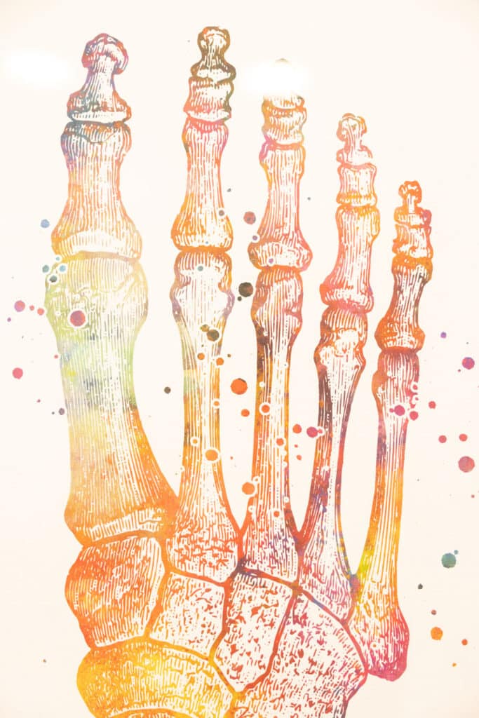
Our feet are made up of three main types of bones: tarsals at the back, metatarsals in the middle, and phalanges, which are the toe bones. Among the tarsals, there are usually seven bones, including the navicular. This bone sits between the ankle bone and the smaller tarsal bones, forming a crucial part of our foot structure.
Understanding this basic foot anatomy sets the stage for grasping the role of the accessory navicular in our overall foot health.
What's an Accessory Navicular?
The human foot is a complex structure composed of various bones, and anomalies can sometimes occur, leading to discomfort and pain.
One such condition involves the presence of an accessory navicular. The accessory navicular is an additional bone that can be found along the inner center arch of the foot. Approximately 2.5 percent of individuals are born with this extra bone, though it often goes unnoticed during early childhood. While not all accessory navicular bones cause discomfort, as individuals grow and engage in physical activities, the accessory navicular may become more prominent, potentially causing discomfort or other foot-related issues, particularly during activities such as walking or engaging in sports.
In some cases, medical attention may be required to address any symptoms or complications associated with this anatomical variation. In this article, we will delve into the intricacies of the accessory navicular and explore the Kidner Procedure, a surgical solution designed to alleviate the associated discomfort.
Why you can trust us:
We believe in the power of specialization. What that means is that we have multiple orthopaedic ankle and foot specialists who treat foot and ankle orthopaedic injuries daily.
We have Ankle and Foot Physical Therapy - our orthopaedic ankle and foot team sees it every week, so you can trust the care you'll receive at POA.
What Does it Feel Like to Have an Issue With an Accessory Navicular
Having this extra foot bone commonly goes noticed. So how can you tell if your foot pain may be related to an accessory navicular? Below is a list of the symptoms you could be experiencing and a list of at-home treatments you can use to try to reduce your discomfort while you wait to see a foot specialist.
Symptoms That Could Mean You Have an Accessory Navicular
Persistent pain or discomfort along the inner arch of the foot
Swelling or tenderness in the affected area
Difficulty wearing certain types of shoes due to irritation
Pain exacerbated by physical activities or prolonged periods of standing
Visible prominence or bump on the inner side of the foot
Limited range of motion in the affected foot
Redness or warmth around the accessory navicular site
Gradual onset of symptoms, which may become more pronounced over time
Discomfort that may interfere with daily activities and physical pursuits
Potential development of flat feet or changes in foot arch structure
How to Reduce Irritation From an Accessory Navicular
Rest: Give your foot a break and avoid activities that exacerbate the pain.
Ice: Apply ice to the affected area for short periods to help reduce inflammation.
Elevation: Elevate your foot when resting to minimize swelling.
Over-the-Counter Pain Medications: Non-prescription pain relievers like ibuprofen or acetaminophen may help manage pain.
Supportive Footwear/Orthotics: To reduce pressure on the accessory navicular, wear shoes with good arch support and cushioning. Consider over-the-counter or custom orthotic inserts to provide additional support and alleviate discomfort.
Modify Activities: Steer clear of high-impact activities that strain the feet excessively, such as running or jumping. Adjust your daily activities to minimize standing or walking for extended periods until you can consult with a specialist.
Why Does an Extra Foot Bone Cause Pain?
When you have an accessory navicular, it can sometimes cause trouble for a tendon in your foot called the posterior tibial tendon. This tendon helps support your arch and foot movement. But with the extra bone hanging around, it might rub against the tendon, causing irritation and, over time, persistent pain. This kind of discomfort can really mess with your day-to-day activities and make it harder to enjoy physical stuff like walking or playing sports. So, it's not just a small thing – it can genuinely affect how you go about your daily routine and stay active.
What is the Kidner Procedure - A Solution for Pain Relief
If you're dealing with pain because of that extra bone in your foot (the accessory navicular), there's a solution called the Kidner Procedure that can really help. This is a surgical fix that focuses on getting rid of the extra bone, which, in turn, can ease the pain you've been feeling. By taking out that pesky extra bit, the procedure aims to bring relief and let you get back to using your foot the way you're used to. It's like a key to kick that discomfort out and help you regain your normal foot function.
The Surgical Process
What is The Kidner Procedure?
The Kidner Procedure is a surgical procedure that begins by separating the accessory navicular from the posterior tibial tendon. After successfully isolating the extra bone, the surgeon proceeds to remove it from the foot altogether. Once the accessory navicular has been excised, the posterior tibial tendon is reattached to the appropriate navicular bone, restoring the structural integrity of the foot.
How is The Kidner Procedure Done?
To perform this procedure, the surgeon typically makes a small incision on the side of the foot, ensuring precision and minimal disruption. This incision serves as the gateway for the surgical maneuvers. After the entire procedure is completed, the incision is carefully closed with stitches, allowing for proper healing. It's a meticulous process aimed at addressing the root cause of the discomfort and ensuring that the foot can heal and function optimally after the surgery.
Recovery & Rehabilitation
What to Expect After The Kidner Procedure
After the Kidner Procedure, the road to recovery usually takes about six weeks. During this time, it's common for patients to rely on crutches to take the weight off their healing foot.
This initial phase is super important because it gives your foot the time it needs to heal up properly. As the recovery moves along, you should start feeling a lot better, with a noticeable drop in the pain that the accessory navicular was causing.
It's like hitting the reset button on your foot troubles, and by the end of those six weeks, you'll likely be well on your way to walking and moving around more comfortably.
The Kidner Procedure stands as a beacon of hope for individuals grappling with persistent foot pain attributed to the accessory navicular. By understanding the intricacies of this condition and the surgical solution offered by the Kidner Procedure, individuals can make informed decisions about their healthcare, seeking lasting relief and an improved quality of life. If you think you are potentially suffering discomfort from an extra foot bone and want to consult with our specialists, you can send us a message online, call us: (609) 924-8131, or text us: (609) 293-2816; We are here to help you get back on your feet.
What is Turf Toe?
Turf toe, a prevalent injury plaguing athletes engaged in dynamic sports like football, soccer, and basketball, arises from the hyperextension of ligaments around the big toe joint, particularly on unforgiving surfaces such as artificial turf or hard grounds. This comprehensive guide navigates through the intricacies of turf toe, elucidating its causes, symptoms, diagnosis, treatment options, and preventive measures. As we unravel the nuances of this common athletic ailment, athletes and sports enthusiasts will gain valuable insights into understanding, managing, and, importantly, preventing turf toe for an uninterrupted and active sporting lifestyle. We will guide you through the intricacies of turf toe, covering its causes, symptoms, diagnosis, treatment options, and preventive measures.
Why you can trust us:
We believe in the power of specialization. What that means is that we have multiple orthopaedic ankle and foot specialists who treat foot and ankle orthopaedic injuries daily.
We have Ankle and Foot Physical Therapy- our orthopaedic ankle and foot team sees it every week, so you can trust the care you'll receive at POA.
Turf toe transpires when the big toe joint undergoes forceful hyperextension, causing ligaments to stretch or tear.
Athletes engaging in sports featuring sudden movements, stops, and turns are especially prone to this injury, impacting the plantar plate beneath the big toe.
Symptoms include pain, swelling, bruising, and instability at the base of the big toe, making walking and bending challenging.
Causes of Turf Toe
Understanding what causes turf toe is crucial for athletes.
It results from intense, abrupt motions, such as forceful upward heel movement beyond designed limits during activities like quick turns, stops, and running.
The toes and ankles endure immense pressure, leading to overstretching or tearing of the plantar plate.
Symptoms of Turf Toe
Recognizing turf toe symptoms aids in timely intervention. Common signs encompass:
Pain on hard surfaces
Swelling
Bruising
Instability at the big toe's base
Decreased toe movement
Difficulty bearing weight on the affected foot.
Diagnosis of Turf Toe
Accurate diagnosis ensures effective treatment. Our physicians conduct a detailed consultation, which may include the following:
A physical examination
Order imaging tests, including X-rays or MRIs, to assess the injury's severity
A personalized and comprehensive treatment plan for your condition and goals
Meet Our Specialists
This blog post is meant to be informative and should not act as a self-diagnosis tool. If you’d like to see one of our doctors, please contact us here.
Turf Toe Treatment
Effective turf toe treatment involves a multi-faceted approach
Rest and Ice Therapy: Resting the foot and applying ice to alleviate inflammation.
Compression and Elevation: Using compression bandages for swelling and elevating the foot.
Pain Medications: Nonsteroidal anti-inflammatory drugs (NSAIDs) provide pain relief.
Footwear and Orthotic Inserts: Wearing stiff-soled shoes with orthotic inserts reduces strain.
Surgery (in severe cases): Surgical intervention may be considered for significant injuries.
Preventing Turf Toe
While prevention can be challenging, athletes can minimize risks by:
Turf toe, a common injury with roots in forceful athletic movements, demands prompt attention. Employing rest, ice therapy, compression, and elevation can mitigate symptoms. Additionally, wearing appropriate footwear and engaging in muscle-strengthening activities are crucial for prevention. If you suspect turf toe, consult with the experts at Princeton Orthopaedic Associates for precise diagnosis and tailored treatment plans. Discover relief and regain your active lifestyle with our dedicated team.
“We are active members in the community, so it is not uncommon for us to see our patients at the grocery store, at school meetings, or in the gym. We take pride in serving them, because when we do, we know we’re doing our part to take care of our community.” - Dr. Jon Ark
Meet Our New Specialist: Dr. Levi - Your Expert in Foot and Ankle Care
We are thrilled to introduce the latest addition to our team of podiatrists, Dr. Levi, a highly skilled specialist in foot and ankle care. Dr. Levi brings a wealth of knowledge, experience, and a strong commitment to patient well-being, making her an invaluable asset to our practice. Whether you're experiencing foot and ankle issues or seeking expert guidance, Dr. Levi is here to provide you with exceptional care.
Educational Excellence
Dr. Levi's journey to becoming a renowned podiatric physician began with her undergraduate education at Rutgers University. Her thirst for knowledge and dedication to healthcare led her to Temple University School of Podiatric Medicine, where she earned her Doctor of Podiatric Medicine degree in 2019.
Residency and Surgical Expertise
To further hone her skills and expertise, Dr. Levi completed an intensive four-year surgical residency program in foot and ankle surgery at Temple University Hospital, a level-1 trauma center known for its commitment to excellence. Her dedication and exceptional performance during her residency were recognized as she was voted chief resident by her peers.
Exceptional Opportunities
During her residency training, Dr. Levi had the privilege of serving as part of Temple's Limb Salvage team, where she played a vital role in preserving patients' limbs and improving their quality of life. She also had the honor of rotating through the prestigious Rubin Institute of Advanced Orthopedics in Baltimore, MD, gaining exposure to a wide variety of rare foot and ankle pathologies.
Passion for Education and Community
In addition to her clinical work, Dr. Levi obtained an associate professorship at Temple University School of Podiatric Medicine. This role allowed her to give back to student and resident education, reflecting her belief that community engagement and education are at the core of being a physician.
Your Path to Better Foot and Ankle Health
Dr. Levi is dedicated to providing compassionate care and innovative solutions to address your foot and ankle concerns. Whether you're dealing with common podiatric issues, complex pathologies, or simply seeking guidance on maintaining optimal foot and ankle health, Dr. Levi's expertise is at your service.
You can learn more about Dr. Levi here. Don't hesitate to contact our office today to schedule your appointment with Dr. Levi. We look forward to helping you achieve better foot and ankle health.
Welcoming Dr. Horling: Your Expert in Podiatric Medicine
We are delighted to introduce Dr. Horling, a highly skilled specialist in podiatric medicine. Dr. Horling joined our team earlier this year. Her impressive background and two decades of experience make her an invaluable asset to our practice. Whether you need foot care, treatment for sports injuries, or wound care, Dr. Horling is here to provide you with exceptional care.
Educational Excellence
Dr. Horling's journey to becoming a renowned podiatric physician began with her undergraduate education at Ursinus College, where she earned a Bachelor of Science Degree in Biology. Her passion for healthcare and dedication to improving patients' lives led her to the Temple University School of Podiatric Medicine, where she obtained her Doctor of Podiatric Medicine Degree in 2000.
Residency Training
To further enhance her skills and expertise, Dr. Horling completed a rigorous three-year residency program at Frankford Hospital (now Jefferson Health) in Philadelphia, PA. This comprehensive program focused on medicine for the first year and then two years of intensive foot and ankle surgery training. Her dedication and commitment to excellence set her on a path to becoming double board certified.
Board Certifications
Dr. Horling's commitment to the highest standards of care is demonstrated through her double board certifications. She is certified in Foot Surgery by the American Board of Foot and Ankle Surgery and in Podiatric Primary Care by the American Board of Multiple Specialties in Podiatry. These certifications reflect her dedication to providing comprehensive podiatric services.
Active Engagement in Podiatric Community
Dr. Horling is an active member of the American Podiatric Medical Society and the New Jersey Podiatric Medical Society. She currently serves on the Board of the NJPMS as the secretary for the Capital Division, further highlighting her commitment to advancing the field of podiatry.
Experience You Can Trust
With over 20 years of practice, Dr. Horling has a wealth of experience treating patients with various conditions, including podiatric medicine, sports injuries, and wound care. Her expertise was further showcased during her tenure as a panelist at the RWJ Wound Care Center in Hamilton for an impressive ten years.
Your Path to Better Foot Health
If you're seeking compassionate care and advanced solutions for your foot and ankle needs, Dr. Horling is here to provide you with exceptional treatment. Don't hesitate to contact our office today to schedule your appointment with Dr. Horling. You can see Dr. Horling's bio here. We look forward to helping you achieve better foot health and overall well-being.