Understand your diagnosis and all treatment options.
Review the benefits, risks, and alternatives with your provider.
Follow all preoperative instructions regarding medications, diet, and fluid intake.
Be aware of what happens during the procedure and the safety checks involved.
Arrange for a ride home and prepare your space for a comfortable recovery.
Begin gentle movement and prescribed exercises on schedule.
Call your provider immediately if you experience any warning signs.
Understanding Your Knee Pain
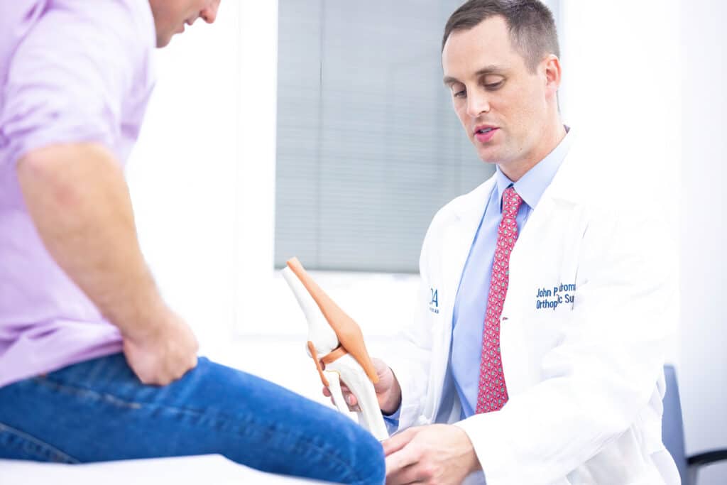
Your knees support you through every step, bend, and climb. Over time, injuries or the natural wear of daily life can lead to pain, swelling, or a feeling of instability. If these symptoms begin to limit the activities you love, your orthopaedic surgeon may suggest knee arthroscopy, a minimally invasive procedure that uses small incisions to look inside the joint and treat the problem.
How the Knee Joint Works
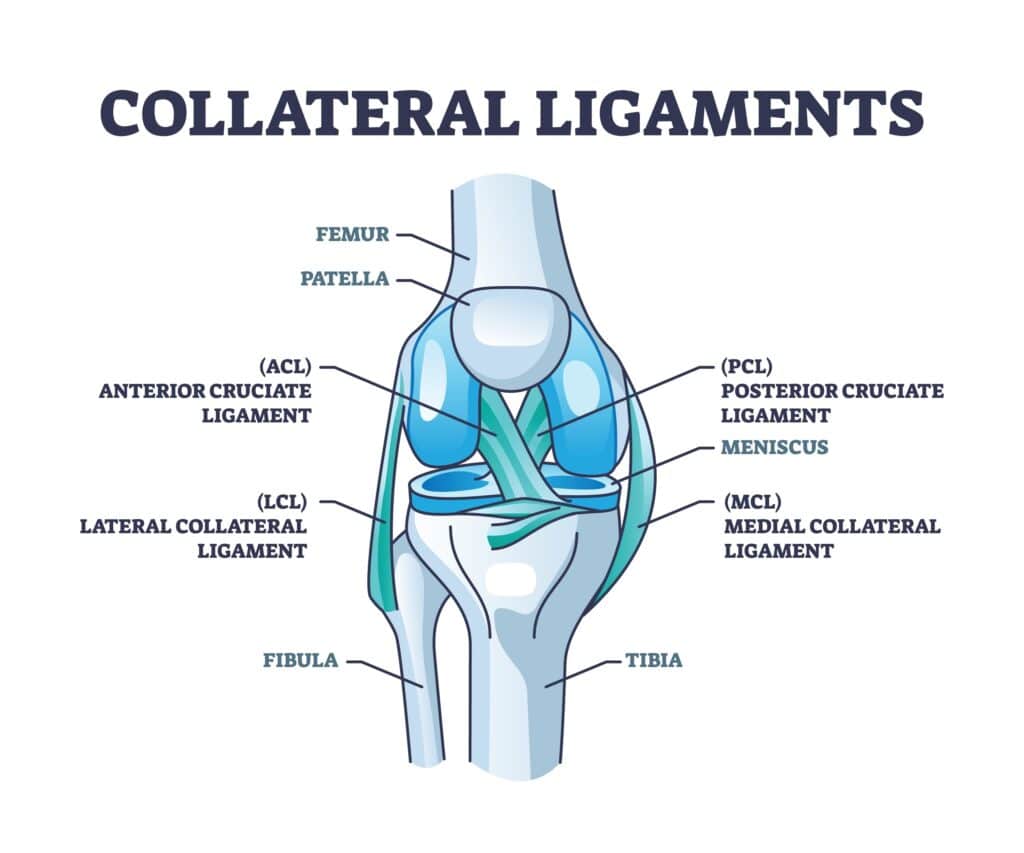
The knee is a complex hinge joint where your thigh bone (femur) and shin bone (tibia) meet. A C-shaped cushion of cartilage called the meniscus sits between these bones to absorb shock. The ends of the bones, along with the back of the kneecap (patella), are coated with a smooth, slick surface called articular cartilage, which allows the joint to glide comfortably. A network of muscles, tendons, and ligaments surrounds the joint to provide strength and stability.
Is Knee Arthroscopy Right for You?
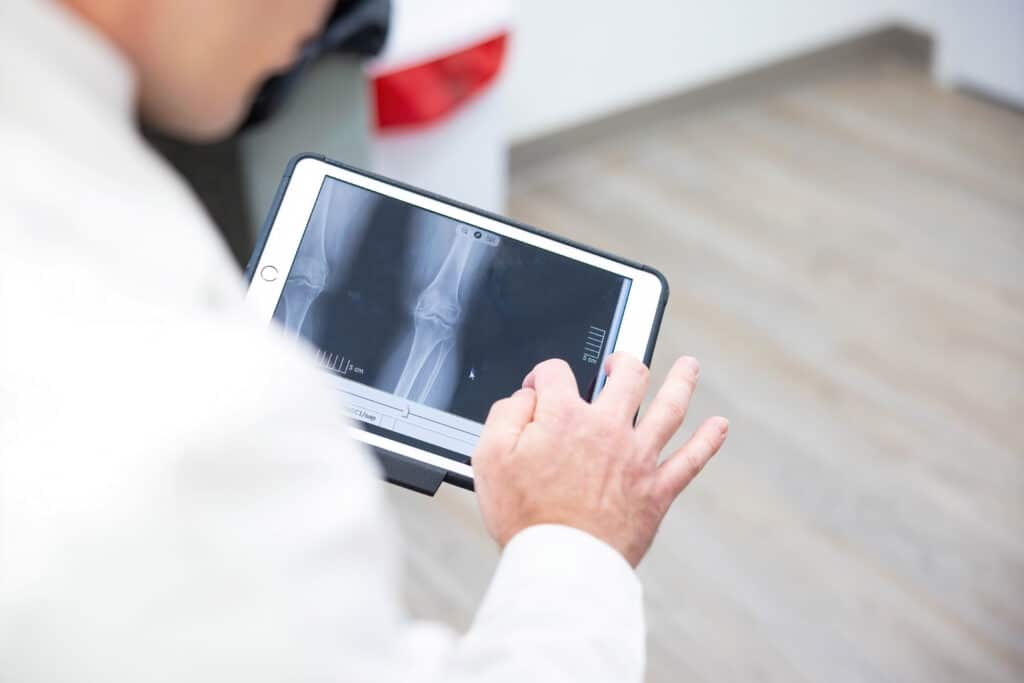
To get an accurate diagnosis, your care team may order imaging tests like an X-ray or an MRI (magnetic resonance imaging). These tests can reveal issues in the soft tissues like ligaments and cartilage, as well as in the bones themselves. Arthroscopy can then be used to confirm what the images suggest and often allows your surgeon to treat the issue during the same procedure.
You and your provider will review the expected benefits and possible risks of arthroscopy. You will also discuss alternatives, which may include medications, physical therapy, activity changes, or wearing a brace. Be sure to ask every question on your mind so you feel informed and confident in your decision.
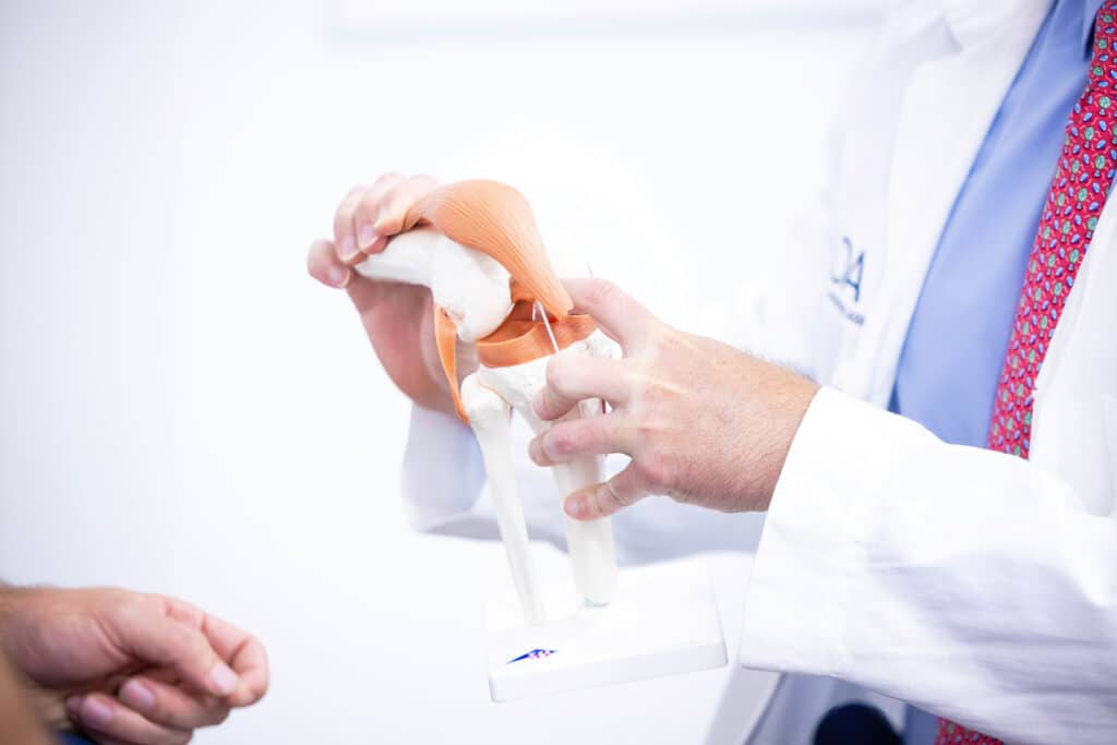
Your Knee Arthroscopy Procedure
What to Expect on Surgery Day
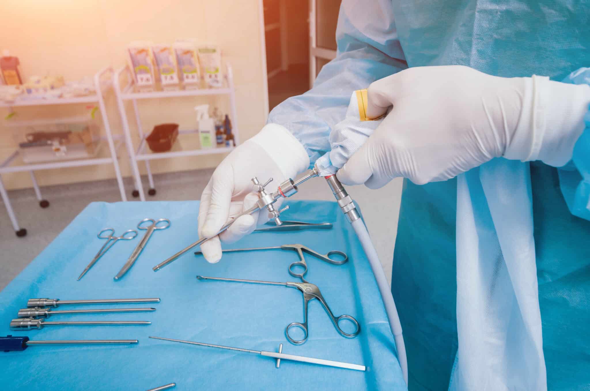
The Procedure: Arthroscopy allows the surgeon to see inside your knee using a thin, lighted camera called an arthroscope. The camera sends live images to a monitor, which guides the precise surgical tools inserted through separate small incisions. This approach allows the surgeon to diagnose and treat many knee problems through incisions that are typically less than an inch long.
Preparing: Before the procedure, inform your surgeon about all the medications and supplements you are taking. You may need to stop some of them ahead of time. You will also be instructed to stop eating and drinking for a set period to ensure your stomach is empty for anesthesia.
The Day of Surgery: On the day of surgery, your team will perform several routine safety checks, including marking the correct knee and confirming your identity and the planned procedure. Anesthesia will keep you comfortable and free from pain. The surgeon then makes two or three small incisions (called portals), places the scope through one, and inserts instruments through the others. Sterile fluid is used to gently expand the joint, improving visibility and helping the surgeon work accurately.
Risks and Possible Complications
Knee arthroscopy is a widely used and very safe procedure. However, like any surgery, it carries some risks. These may include:
Bleeding, infection, or blood clots
Stiffness or ongoing knee pain
Injury to blood vessels, nerves, or skin around the knee
Damage to cartilage, the meniscus, or ligaments
The need for additional surgery
Other specific risks as discussed by your surgeon
Common Conditions Treated with Arthroscopy
Arthroscopy can address a variety of common knee problems:
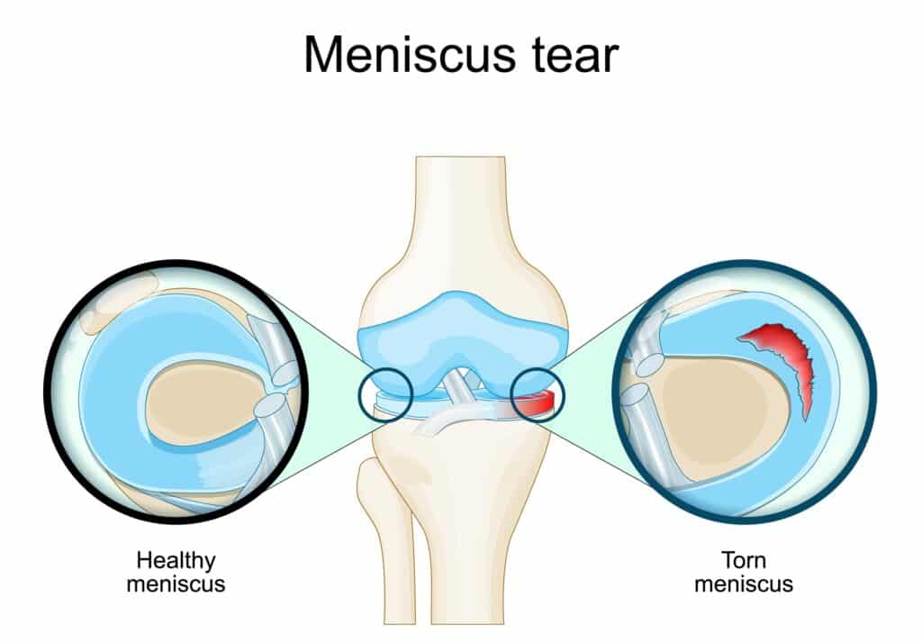
A Healthy Knee
The Problem: Repeated squatting or a sudden twist can tear the meniscus. This may cause pain or swelling, and your knee may catch or lock when you move.
The Solution: Torn tissue on the inner portion of the meniscus is often trimmed away (a meniscectomy). Tears near the outer edge, which has a better blood supply, may be repaired with sutures.
Meniscus Tears
The Problem: Repeated squatting or a sudden twist can tear the meniscus. This may cause pain or swelling, and your knee may catch or lock when you move.
The Solution: Torn tissue on the inner portion of the meniscus is often trimmed away (a meniscectomy). Tears near the outer edge, which has a better blood supply, may be repaired with sutures.
ACL Tear
The Problem: A sudden pivot, cut, or awkward landing can tear the ACL. Patients often report a “pop,” rapid swelling within hours, and instability with pivoting or cutting.
The Solution: Low-demand or partial tears may be managed with structured rehab and bracing. Symptomatic complete tears, especially in patients returning to sports, are typically treated with arthroscopic ACL reconstruction using a tendon graft; primary ACL repair is reserved for select tears.
Cartilage Wear
The Problem: Articular cartilage can wear down, and loose pieces can float inside the joint. You may notice pain, stiffness, or a grinding sensation.
The Solution: The surgeon can remove loose fragments that irritate the joint to reduce catching and pain, and smooth down damaged cartilage surfaces.
Patella (Kneecap) Issues
The Problem: Articular cartilage can wear down, and loose pieces can float inside the joint. You may notice pain, stiffness, or a grinding sensation.
The Solution: The surgeon can remove loose fragments that irritate the joint to reduce catching and pain, and smooth down damaged cartilage surfaces.
Your Recovery at Home
Immediately After Surgery
When the procedure is finished, your small incisions will be closed and covered. In the recovery room, your knee will be bandaged, iced, and elevated to limit swelling. You will receive pain medication and be monitored by a nurse until it is safe to go home. Anesthesia and pain medicine can make you drowsy, so you must arrange for an adult to drive you home.
Caring for Your Knee
Follow your surgeon’s instructions carefully to ensure a smooth recovery. Key steps include:
Elevate your leg above the level of your heart as much as possible to reduce pain and swelling.
Ice your knee for 20-30 minutes several times a day for the first few days. Wrap the ice pack in a thin towel to protect your skin.
Keep incisions dry. Shower only when your provider says it is safe, and cover your leg with plastic to keep the bandages dry.
Manage weight-bearing. You may go home with crutches. Follow your weight-bearing directions carefully so your knee can heal properly.
Exercises for a Strong Recovery
Oftentimes, physical therapy is prescribed. But at home, you can work on many of the following.
Gentle movement is critical for healing. Your provider or physical therapist will guide you through a plan to restore motion and strength. Start these simple motions as soon as you are told it is safe to improve blood flow and help prevent blood clots.
Ankle Pumps: Point your foot down, then flex it up. Move your foot in circles several times throughout the day.
Quadriceps Sets: While lying down, tighten the muscles on the front of your thigh and press the back of your knee toward the surface. Hold for 5 to 10 seconds, then relax. Repeat as directed.
Straight Leg Raises: Lying down, keep your knee straight and lift your leg 8 to 12 inches off the surface. Hold for 5 seconds, then lower slowly. Repeat as directed.
When to Call Your Provider
Contact your provider’s office right away if you notice any of the following:
Fever of 100.4° F (38° C) or higher
Pain that does not improve with medication and rest
Swelling that does not improve with elevation and icing
Increased redness, warmth, or drainage from the incision sites
Bleeding that soaks through your bandages
New or worsening numbness in your leg or foot
Severe nausea or vomiting
Returning to Your Activities
Recovery time varies based on your specific procedure, the condition of your knee, and your overall health. Many people with desk jobs can return to work in about one week. Jobs that require prolonged standing or heavy activity may require more time off. With consistent effort in your rehabilitation, most people can return to their normal active lifestyle within one to two months.
Knee arthroscopy is a powerful tool for diagnosing and treating the cause of your knee pain. By preparing as instructed and following your post-operative plan closely, you give yourself the best chance of returning to your activities with less pain and improved function.
This blog post is meant to be informative and should not act as a self-diagnosis tool. If you’d like to see one of our doctors, please contact us here.
Pain On the Outside of Your Knee Could be IT Band Syndrome.
You don’t have to be a marathon runner to feel that nagging ache on the outside of your knee. The important thing? It might not actually be your knee. It might be a tight IT band, and unlike joint injuries, it requires a different kind of treatment focused on mobility and muscle balance.
Maybe it starts during your daily walk, or when you’re going up stairs. Perhaps it flares up when you get up from your desk or out of the car. It might even wake you up at night, pulsing in your outer thigh or hip, making it impossible to get comfortable. It doesn’t feel like an injury yet, the pain keeps coming back.
If this sounds familiar, there’s a good chance your iliotibial band (IT band) is involved. And the condition you might be dealing with is called IT Band Syndrome, a common cause of outer knee and hip pain that affects far more than just athletes.
Let’s walk through what’s happening in your body, why it hurts, and most importantly, what you can do to start feeling better.
Here Are The Things You Need to Know About IT Band Syndrome
What You Should Know:
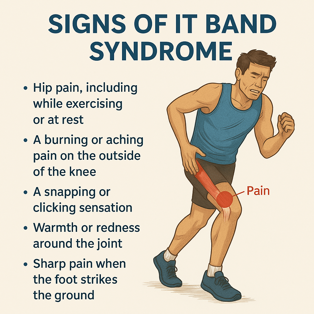
Pain on the outside of your knee or thigh is a common sign of IT Band Syndrome, and it's not just a runner's problem.
You don’t have to be an athlete. Everyday movement, prolonged sitting, or climbing stairs can all trigger symptoms.
The pain is caused by tightness and friction where the IT band rubs against bone near the knee.
Stretching the IT band itself won't solve the issue. Instead, focus on loosening surrounding muscles and improving strength.
Symptoms often show up with walking, going downstairs, or after long periods of inactivity.
The discomfort may travel up to your hip, outer thigh, or glute region, especially when lying on your side.
Causes often include weak glutes or core, poor posture, or worn-out shoes that don’t offer enough support.
Foam rolling can help, but you want to target the glutes, quads, and TFL, not directly on the IT band.
Recovery requires more than rest. Strengthening, mobility work, and correcting how you move are essential.
Seeing a physical therapist can help you address the root cause and get long-term relief.
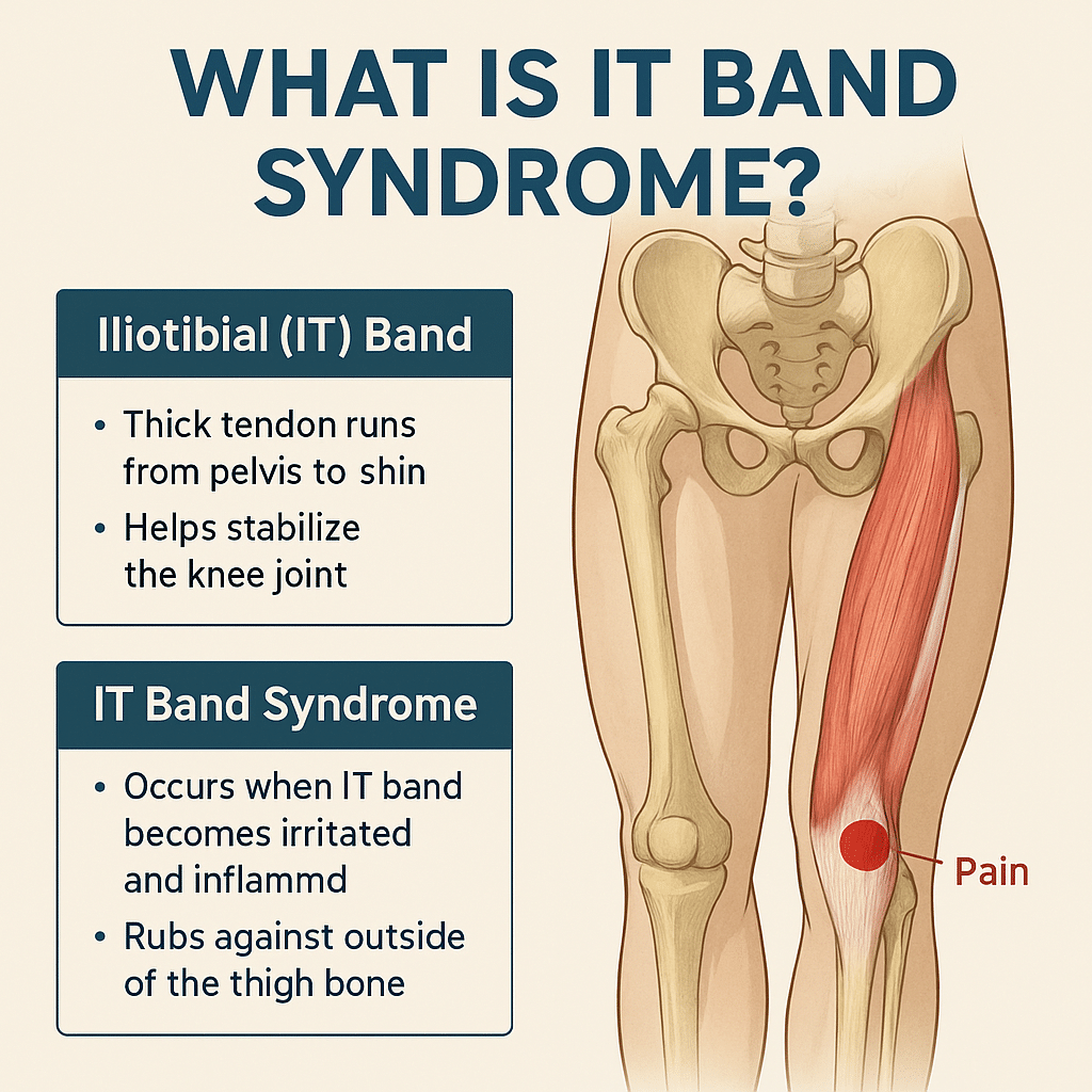
What Is the IT Band and Why Does It Get Tight?
The iliotibial (IT) band is a thick, fibrous band of connective tissue that runs down the outside of your leg, from your hip to just below your knee. Think of it as a support strap that helps stabilize your knee and assist with hip movement.
When the IT band gets too tight, often due to repetitive movement, muscle imbalances, or poor posture, it can rub against the bone at the outer knee. This creates irritation, inflammation, and pain, commonly known as IT Band Syndrome (ITBS).
And while it’s often associated with athletes, it’s just as common in walkers, desk workers, parents, nurses, retail workers, and anyone who’s on their feet a lot, or not enough.
What Does IT Band Syndrome Feel Like?
Here are common, real-world symptoms of IT Band Syndrome in everyday life:
Aching or burning pain on the outside of the knee
Tightness or pulling in the outer thigh
Pain or discomfort going up or down stairs
Sharp pain with walking or standing for extended periods
Tenderness at the hip or discomfort when lying on your side
Clicking or snapping near the hip or knee
These symptoms often start mild, but become more consistent if left unaddressed.
What Causes IT Band Syndrome in Non-Athletes?
Even without intense training, everyday habits can contribute to ITBS:
Sitting for long periods without movement
Poor posture or weak hip/core muscles
Uneven walking surfaces (like sloped sidewalks or hilly neighborhoods)
Wearing worn-out or unsupportive shoes
Standing or walking with one leg favored over the other
Repetitive daily movement without adequate strength or flexibility
What Causes IT Band Syndrome in Athletes?
While the core problem is the same (tightness and friction along the IT band), athletes often develop ITBS due to training volume and biomechanics. Common athletic triggers include:
Sudden increases in mileage or intensity (especially in runners and cyclists)
Downhill running or running on sloped surfaces
Repetitive activities involving knee flexion and extension
Weakness in hip abductors, glutes, or core stabilizers
Poor running form or gait asymmetries
Worn shoes or improper footwear for training conditions
Overtraining without proper rest and recovery
IT Band Syndrome is common among:
Distance runners
Cyclists
Soccer and hockey players
HIIT or CrossFit athletes
Skiers or hikers tackling long descents
💡 Tip for athletes:
Strengthen your hips and glutes, cross-train, and make sure your recovery matches your training load
.
Why Is IT Band Pain So Persistent?
The IT band isn’t a muscle, it’s actually connective tissue. That means:
You can’t stretch it the same way you stretch a muscle
If surrounding muscles (like glutes and hip flexors) are tight or weak, the IT band picks up the slack
Without correcting imbalances, foam rolling or resting alone won’t fix it
Over time, the friction and inflammation can become chronic and much harder to treat.
How Is IT Band Syndrome Treated?
Treatment focuses on reducing inflammation, improving mobility, and correcting muscle imbalances.
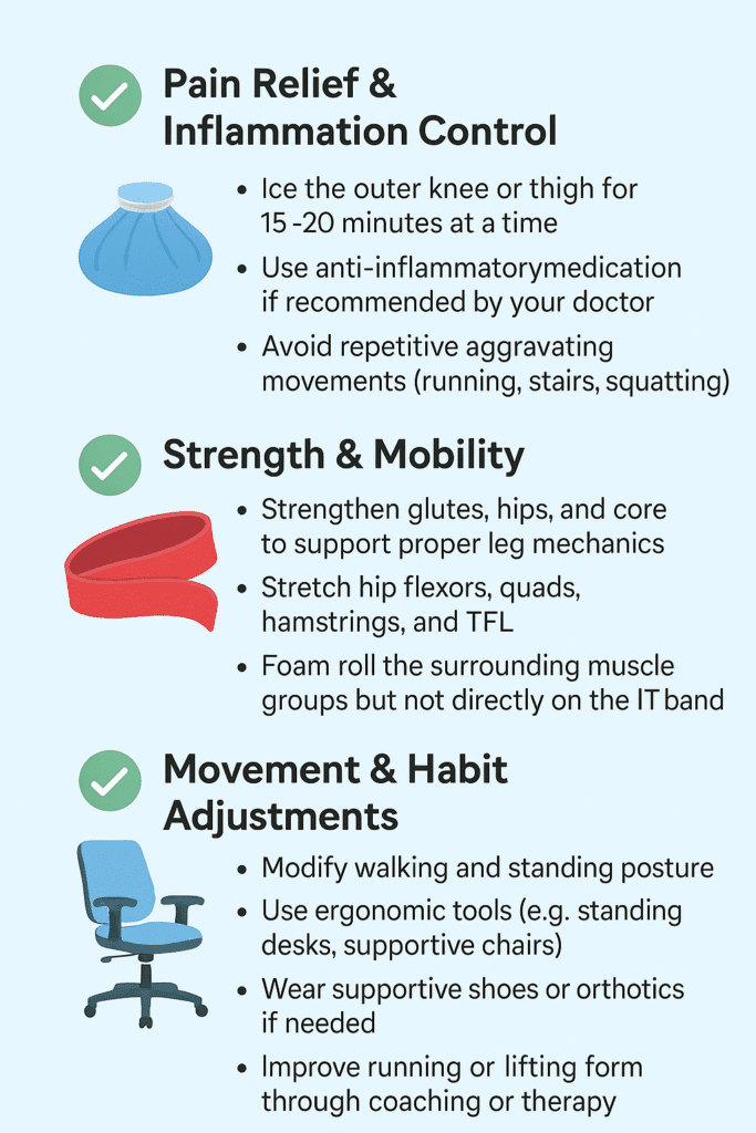
✅ Pain Relief & Inflammation Control
Ice the outer knee or thigh for 15–20 minutes at a time
Use anti-inflammatory medication if recommended by your doctor
Strengthen glutes, hips, and core to support proper leg mechanics
Stretch hip flexors, quads, hamstrings, and TFL
Foam roll the surrounding muscle groups but not directly on the IT band
✅ Movement & Habit Adjustments
Improve running or lifting form through coaching or therapy
Modify walking and standing posture
Use ergonomic tools (e.g., standing desks, supportive chairs)
Wear supportive shoes or orthotics if needed
How Long Does IT Band Syndrome Take to Heal?
The length of time to recover from IT Band Syndrome depends on how long you've had symptoms and whether you're treating the root cause:
Severity
Recovery Time
Notes
Mild
2-3 Weeks
Rest and stretching may help quickly if caught early
Moderate
4-6 Weeks
Requires active rehab including movement correction
Chronic
2+ Months
Long-standing tightness or inflammation takes time to unwind
Everyday Life with IT Band Syndrome
IT Band Syndrome doesn't just show up during workouts; it can quietly interfere with our daily routine, mobility, and overall comfort. Without treatment, ITBS can impact your:
Ability to walk, climb stairs, or stand comfortably
Sleep (especially side-sleepers)
Workday (especially for those on their feet)
Confidence in movement and balance
Long-term joint health if compensatory patterns develop
And for athletes, it can put your training on pause or create a cycle of recurring injuries.
Should I See a Doctor for a Tight IT Band?
If you have been experiencing symptoms of IT Band Syndrome and you haven't found relief, you should consult with a specialist. Especially if:
The pain has lasted more than a week
You’ve tried rest but symptoms return
You’re changing how you move to avoid pain
You’re unable to stay active or complete daily tasks comfortably
At Princeton Orthopaedic Associates, we have physicians from multiple specialties that can help you get to the root of your tight IT band and help set you off on the path to recovery.
- A physical therapist is often the next step after diagnosis for hands-on treatment and long-term recovery.
Our specialists will identify the root cause of your tightness, guide you through targeted corrective exercises, and help you improve how you move—not just mask the symptoms.
Stop Living With IT Band Pain
Whether you're training for a race or just trying to get through the workday without pain, IT Band Syndrome can be disruptive, but it's absolutely treatable. The key isn’t just stretching or resting, it's understanding why the IT band is tight and retraining your body to move in a healthier, more balanced way.
This blog post is meant to be informative and should not act as a self-diagnosis tool. If you’d like to see one of our doctors, please contact us here.
Pain After A Pop In The Knee
An ACL tear typically occurs during sudden pivoting, awkward landings, or stops, which are common in sports such as basketball, soccer, and skiing. It usually starts with a moment, an awkward pivot during a pickup basketball game, a sudden stop on the soccer field, or landing just slightly wrong after a jump. You might feel a sharp pain, instability, or hear that telltale pop. You go down, maybe hoping it’s nothing, but your knee swells, and walking becomes difficult. That’s the moment many athletes, professional, weekend warriors, or even teenagers, begin their journey with an ACL tear.
Understanding how to recognize a minor knee issue needing minimal home treatment versus what may be an ACL tear can be critical to the proper treatment and the fastest path to healing.
What Is the ACL and Why Does It Matter?
Your knee is one of the most complex joints in your body, and the ACL is one of its most important components. It plays a huge role in keeping your knee stable and allowing you to move with confidence, whether you're sprinting down a field or simply walking downstairs.
The ACL (anterior cruciate ligament) is one of the four major ligaments in your knee, connecting your thigh bone (femur) to your shinbone (tibia). Its job is to stabilize the knee, especially during rotation, pivoting, and rapid direction changes. That makes it crucial for athletes, but also important for anyone who walks, runs, or climbs stairs.
When the ACL tears, it doesn’t heal on its own. And unlike muscles, ligaments don’t regenerate well without surgical reconstruction.
ACL Tear Symptoms: What You Might Experience
If you’ve injured your knee and are wondering if it’s your ACL, you’re not alone. Knowing what symptoms to look for can help you decide whether it’s time to see a doctor or get imaging.
Here’s what people often report:
A “popping” sound or sensation at the moment of injury
Immediate pain, sometimes severe enough to stop activity
Swelling that begins within a few hours
Instability or “buckling”, especially when trying to pivot or walk
Loss of full range of motion
Some people can walk after an ACL tear, especially once swelling subsides, but the knee often feels unstable. Grade 1 (mild) tears may feel like soreness and instability under stress, but they’re rare. By two weeks post-injury, swelling may reduce, but instability often persists.
Grade 3 (Complete tear): Surgical (ACL reconstruction) 6–12 months before full return to sport
MCL Tear Recovery:
Grade 1 (mild, stretching without tearing): 1–3 weeks
Grade 2 (moderate, partial tear): 3–6 weeks with bracing and PT
Grade 3 (complete): 6–10 weeks, may involve bracing or rare surgical repair
Why you can trust us:
We have multiple highly specialized, board-certified, fellowship-trained orthopaedic surgeons.
We know that we serve people - actual humans - not random orthopaedic conditions. That drives us to compassionate care.
The world of orthpaedics is constantly evolving. Our orthopaedic surgeons are constantly evaluating new techniques, tools and methods to serve our community even better.
We provide outcome-focused treatment. We work with our patients to achieve their goals, all while developing custom treatment plans that fit our patient's lives.
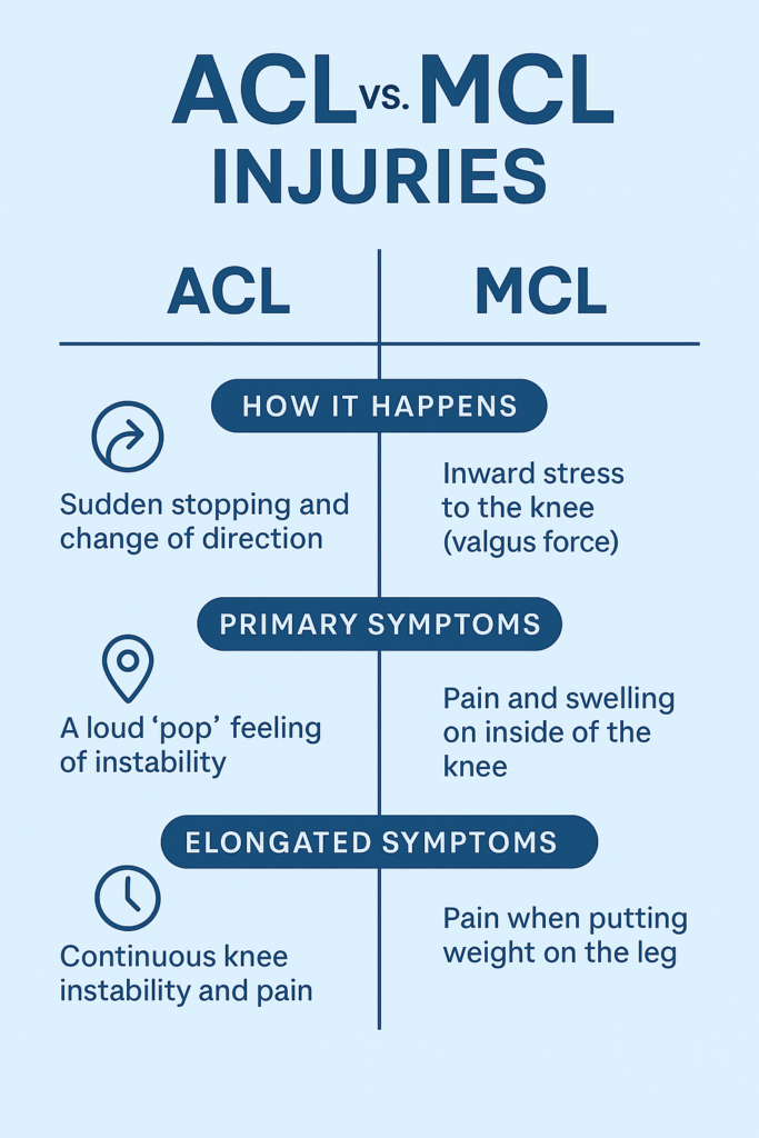
Knee injuries can be confusing because symptoms often overlap. The ACL and MCL are two different ligaments with different functions, injury mechanisms, and treatment approaches. Understanding the difference is critical for proper recovery.
Many people confuse ACL and MCL (medial collateral ligament) injuries. Here’s how an ACL tear and MCL tear differ:
ACL Tear
MCL Tear
Location
Inside the knee, central
Inside of the knee (medial side)
Mechanism
Pivoting, cutting, or landing
Direct blow to outer knee or overstretching
Sound
Often a pop
Less commonly a pop
Swelling
Fast and significant
Less severe swelling
Instability
Knee feels unstable or “gives out”
Usually more stiff than unstable
Healing Potential
Does not heal on its own
Often heals without surgery
First-line Treatment
Physical therapy or surgical reconstruction
Bracing, rest, and physical therapy
Surgery Needed?
Often required in active patients
Rarely required (unless Grade 3 + other injuries)
Return to Sport
6–12 months (after reconstruction)
4–12 weeks (depending on severity)
Key difference: An MCL tear can often heal with rest and bracing. An ACL tear usually won’t.
Can You Tear Both ACL and MCL at Once?
Yes, and this is more common than people think, especially in sports injuries. This is called a combined ligament injury and often involves the ACL, MCL, and/or meniscus. These cases require specialized surgical planning and longer rehabilitation timelines, making early diagnosis even more critical.
How to Know Which One You’ve Injured
While some clues (pain location, swelling speed, mechanism) may point toward one ligament over the other, you cannot reliably self-diagnose an ACL or MCL tear. Some people with a complete ACL tear are still able to walk or bend their knee, which can be misleading.
We recommend consulting one of our sports medicine specialists or an orthopedic knee surgeon as soon as possible. A timely and accurate diagnosis gives you the best chance of a full recovery and of avoiding chronic knee issues.
Adolescent ACL Tear vs. Adult ACL Tear
When it comes to ACL injuries, age matters. Kids and teens are still growing, and that can make treatment more complicated. What’s best for a 14-year-old soccer player may be very different from what’s recommended for a 30-year-old runner.
ACL injuries are increasing in adolescents, especially teenage athletes. The growth plates (areas of developing cartilage near the ends of long bones) in kids add complexity:
Non-surgical approaches may be prioritized in younger teens to avoid damaging growth plates.
Modified surgical techniques (like physeal-sparing procedures) are used if surgery is necessary.
Rehabilitation may need to be longer to protect future growth and return-to-play safely.
In adults, decisions are often based on lifestyle, activity level, and degree of instability.
Surgery isn't always required for an ACL tear, but it can often be recommended. Once you’ve torn your ACL, the big question is: Do you need surgery? The answer depends on your goals, age, activity level, and the nature of the tear. For some, physical therapy may be enough. For others, reconstruction is the most straightforward path back to full function. Your treatment path is specific to you, and our specialists will build a plan that meets the needs of your injury and desired recovery outcome.
Non-Surgical ACL Tear Treatment (select cases)
Mild (grade 1) sprains may recover within 3–6 weeks with rest and rehab.
For:
Low-demand lifestyle (non-athletes)
Partial tears
Older adults
No instability with daily activity
Approach:
Activity modification
Physical therapy to strengthen surrounding muscles (especially quads and hamstrings)
Bracing for certain activities
Surgical ACL Tear Treatment (Reconstruction)
Surgery may sound intimidating, but for many people, it offers the best chance at regaining full knee stability and returning to high-level physical activity. The procedure is common, safe, and continually improving.
Most active individuals, especially athletes or younger patients, choose ACL reconstruction. Here’s how it works:
Torn ACL is replaced with a graft (from your own hamstring, patellar tendon, quadriceps tendon, or a donor).
Surgery is typically minimally invasive (arthroscopic).
Recovery involves 6–12 months of guided physical therapy.
Factors influencing surgery:
Presence of other injuries (meniscus tear, cartilage damage)
Age and activity level
Desire to return to sports
Degree of instability
What Not To Do After an ACL Tear
Sometimes what you don’t do is just as important as what you do. The wrong move after an ACL tear can worsen the injury or lead to complications down the line.
Don’t ignore instability. Repeated “giving out” episodes can damage the meniscus or cartilage.
Don’t rush rehab. Returning to play too soon raises your risk of re-tear (or injuring the other knee).
Don’t skip the MRI. X-rays won’t show ligament damage. An MRI is needed to confirm the tear and check for other injuries.
Don’t rely solely on a knee brace if you're planning to return to sport, it doesn't replace ligament function.
Leaving an ACL tear untreated can lead to further joint damage, including cartilage wear or meniscus tears.
Can You Still Use Your Knee with a Torn ACL?
Some people can still walk, squat, or bend their knee shortly after tearing their ACL. However, without stability, these motions can cause further injury. If you suspect you have an ACL tear we recommend you see a orthopaedic specialist as soon as possible for a comprehensive evaluation.
Life After an ACL Tear: Hope, Patience, and Progress
An ACL tear is a detour, not a dead end. With the right care, commitment, and patience, people of all ages get back to running, jumping, and playing, often even better than before.
Tearing your ACL can feel like the end of your athletic identity, but it’s not. Thousands of people, from high school athletes to weekend hikers to pro players, successfully return to sports and active lifestyles every year.
The key is getting the right diagnosis, choosing the right treatment path for your goals, and committing to smart, structured rehab.
Diagnosis: Why You Need a POA Orthopaedic or Sports Medicine Specialist
While some symptoms can help differentiate between the two, it’s extremely difficult to diagnose knee ligament injuries accurately without imaging and specialist assessment.
Here’s why seeing a POA or orthopedic knee specialist is essential:
Physical tests (like Lachman or valgus stress test) must be performed with skill and interpreted in context.
MRI scans are required to confirm the exact ligament involved and assess associated injuries (meniscus, cartilage).
Misdiagnosis can delay proper healing. For example, treating an ACL tear like an MCL sprain could lead to long-term instability or joint damage.
Bottom Line: Always get a clinical evaluation with a knee specialist, especially if you heard a pop, felt instability, or have swelling. Don’t self-diagnose based on symptoms alone.
Meet Our Orthopaedic Knee Specialists
Can You Recover from an ACL Tear? Here’s What to Expect
Tearing your ACL may feel overwhelming, but it's not the end of your active lifestyle. Whether you're a competitive athlete or someone who just wants to move without fear, recovery is possible with the right approach. From early diagnosis and personalized treatment plans to structured rehab and return-to-play timelines, every step forward matters. Understanding your options is the first step toward getting back to what you love, with strength and confidence.
If you’re reading this, you may be worried about what’s next. Take a breath, you’re not alone. Understanding your injury is the first step toward healing. Now it’s time to take action. If you suspect an ACL tear, don’t wait. Get evaluated by a sports medicine physician or orthopedic specialist. Early diagnosis means earlier healing and a better chance of getting back to doing what you love.
ACL Tear vs. Meniscus Injury: Key Differences at a Glance
While both ACL and meniscus injuries are common in athletes and active individuals, they are very different in structure, symptoms, and recovery needs. Knowing the distinctions can help guide proper diagnosis and treatment.
How an ACL Tear and Meniscus Tear Happen
ACL Tear:
Typically from a sudden pivot, change in direction, or awkward landing
Often non-contact, though can also result from trauma
Common in sports like soccer, basketball, skiing
Meniscus Tear:
Often caused by twisting the knee while the foot is planted
Can occur with or without an ACL tear
May result from degeneration in older adults or a sharp movement in younger athletes
Primary (Immediate) Symptoms
Symptom
ACL Tear
Meniscus Tear
Popping Sound
Very common
May occur, but less dramatic
Swelling
Rapid (within hours)
Gradual (over 24–48 hours)
Instability
Knee may "give out"
Usually feels stable
Pain Location
Deep or central knee
Side or back of knee (depending on tear location)
Mobility
Loss of motion due to swelling and instability
May still walk, but discomfort with twisting/squatting
Prolonged/Chronic Symptoms (If Left Untreated)
Symptom
ACL Tear
Meniscus Tear
Knee Giving Out
Frequent instability, especially during pivoting
Rarely unstable
Locking or Catching
Uncommon
Very common — knee may catch or lock during motion
Grinding or Clicking
Occasionally
Common, especially with movement
Degeneration Risk
Higher if combined with meniscus injury
Increases risk of arthritis over time
Return to Activity
Difficult without surgery for active individuals
Sometimes possible without surgery, depending on severity and tear location
A meniscus tear often presents with joint line tenderness and mechanical symptoms (like locking), while an ACL tear leads to feelings of instability and swelling shortly after injury. However, since both can coexist, and symptoms can overlap, accurate diagnosis with an MRI and specialist evaluation (by a POA or orthopedic physician) is essential. Read more about meniscus tears.
Quick Overview
ACL
Meniscus
Function
Stabilizes the knee
Cushions and supports joint movement
Injury Type
Ligament
Cartilage
Instability?
Yes
Rarely
Locking
Rare
Common
Needs Surgery
Often (for active patients)
Sometimes, depending on tear type
This blog post is meant to be informative and should not act as a self-diagnosis tool. If you’d like to see one of our doctors, please contact us here.
Tweaked your knee, but the pain isn't going away?
You were mid-pivot, chasing a ball or turning to grab something behind you, when a sharp pop hit your knee. Not loud, but distinct. You paused, unsure if it was serious. Maybe just a tweak, you thought. But within hours, the swelling crept in, the joint stiffened, and walking suddenly felt unfamiliar. That small twist? It turned into something much bigger.
That moment likely marked the beginning of a meniscus tear—a common yet disruptive injury affecting the cartilage in your knee. Whether it's from a sudden injury or years of wear and tear, the result is often the same: pain, limited movement, and questions about what comes next.
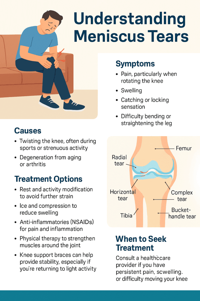
What Is a Meniscus Tear?
Inside each of your knees are two rubbery, wedge-shaped pieces of cartilage: the medial and lateral menisci. These act like shock absorbers between your thighbone and shinbone, helping to distribute weight and stabilize movement. A tear occurs when this cartilage is damaged—usually from twisting motions or degeneration over time.
You don't have to be an athlete for this to happen. A quick squat, an awkward turn, even standing up too fast with pressure on the joint can be enough, especially if the cartilage is already weakened with age.
Common Causes
There are two primary culprits behind a torn meniscus:
Trauma or sudden movement, like twisting or pivoting during sports, or while playing with your kids!
Degeneration, where age-related wear and tear thins and weakens the cartilage, making it easier to tear with minor movements.
Both scenarios are incredibly common. Lifting a heavy box incorrectly or kneeling on a hard surface for too long can be all it takes.
Meet Our Orthopaedic Knee Specialists
Symptoms of a Meniscus Tear
The first few hours after the tear are often the most telling. At first, discomfort may be the only symptom of a meniscus tear you might feel. Or, the only symptoms of a meniscus tear present at first are just a dull, persistent ache, made worse by movement. You might feel fine while sitting, but as soon as you try to walk or bend, your knee doesn't cooperate. Some describe it as a "stuck" sensation, where the joint feels like it won't fully extend or flex without pain or resistance. But then the pain deepens, swelling begins, and your range of motion shrinks even more.
Clicking, popping, or catching during movement can also indicate a torn flap of cartilage catching in the joint. Check out this post to read more about the Types of Meniscus Tears.
Additional Symptoms of a Meniscus Tear:
Locking, catching, or the feeling of instability
Sharp or aching pain, often on the inner (medial) side of the knee
Swelling that develops gradually
A popping sound or sensation during injury
Difficulty straightening or bending the knee
If you're looking for clarification on the symptoms of a meniscus tear, you are not alone. Many people deal with a torn meniscus and don't realize the seriousness until the stiffness and pain don't go away.
Do You Need to See a Doctor For a Meniscus Tear?
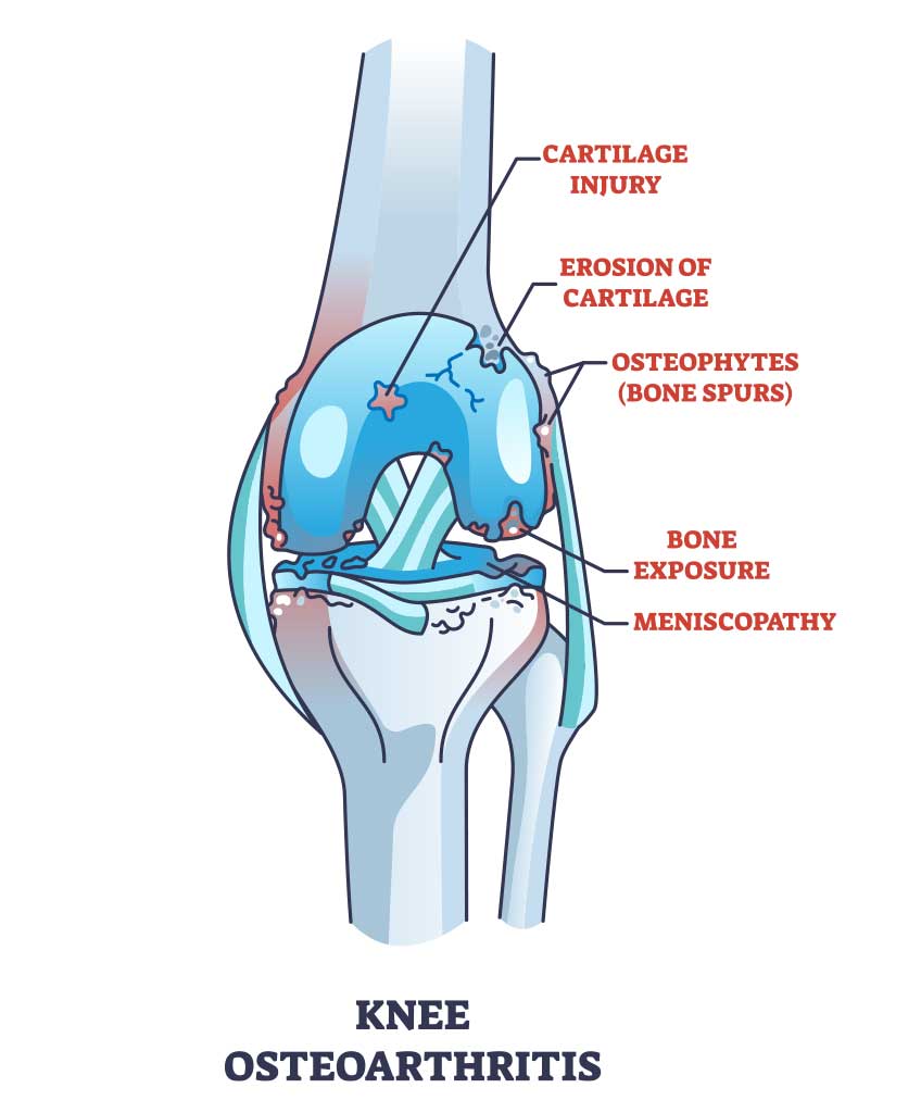
If you're hoping it will just go away, consider this: untreated meniscus tears can worsen over time, leading to more pain and even long-term joint issues like osteoarthritis.
Seek professional help if:
Pain persists beyond a few days
Swelling continues or worsens
You experience locking, buckling, or instability
You can't put normal weight on the leg
Ignoring it risks further tearing or cartilage breakdown. Early diagnosis often means better, less invasive treatment options.
Why you can trust us:
We have multiple highly specialized, board-certified, fellowship-trained orthopaedic surgeons.
We know that we serve people - actual humans - not random orthopaedic conditions. That drives us to compassionate care.
The world of orthpaedics is constantly evolving. Our orthopaedic surgeons are constantly evaluating new techniques, tools and methods to serve our community even better.
We provide outcome-focused treatment. We work with our patients to achieve their goals, all while developing custom treatment plans that fit our patient's lives.
A physical exam can often provide early clues. A clinician will test your range of motion and apply gentle pressure or rotation to identify pain points. In many cases, imaging, like an MRI, is used to confirm the diagnosis and pinpoint the severity and location of the tear.
Meniscus Tear Remedy
Not all meniscus tears require surgery. If you're looking for a meniscus tear remedy, treatment depends on the type of tear, location, and severity of the tear, as well as your activity level and age.
Conservative Meniscus Tear Remedy Approaches:
Rest and Activity Modification
One of the first things you can do is to give your knee a break! If you are able to identify them, avoid movements thatmake the pain worse. Common ones include twisting, squatting, or any high-impact activities. Resting allows the damaged cartilage in your knee to settle and inflammation to subside which gives your body a chance to begin healing. This doesn't mean you have to be totally immoble, but be mindful of your movements and eliminate anything that causes discomfort or strain.
Ice and Compression
Applying ice packs to your knee helps reduce swelling and numbs the area, easing the pain. Aim for 15–20 minutes every few hours in the first few days. Pair this with a compression bandage or sleeve to minimize inflammation and support the knee structure. Together, they help control the body's inflammatory response and provide short-term relief while preventing further irritation.
Anti-inflammatories (NSAIDs)
Over-the-counter medications like ibuprofen (Advil) or naproxen (Aleve) can significantly reduce inflammation and help manage pain. These drugs target the body’s natural inflammatory chemicals, making it easier to move the joint and complete daily activities without aggravating the tear. Always follow dosage instructions and consult your doctor if you’re taking them for more than a few days.
Physical Therapy
Once the pain and swelling are under control, targeted exercises become essential. A physical therapist will guide you through movements designed to strengthen the muscles surrounding your knee—especially the quadriceps and hamstrings. This not only speeds up recovery but also restores joint stability, improves flexibility, and reduces the risk of re-injury.
Knee Support Braces
A well-fitted knee brace offers additional stability, particularly when walking or performing light activities. Braces help limit unwanted lateral movement and protect the joint during recovery. If your knee tends to feel unstable or you're easing back into exercise or work, wearing a brace can provide the support and confidence you need to move safely.
Small tears near the outer edge, where the blood supply is richer, often heal with conservative care.
Surgical Options:
If the tear is large, causes locking, or doesn't improve, arthroscopic surgery may be recommended. Options include:
Meniscus repair (stitching the cartilage back together)
Partial meniscectomy (removing the torn section)
Total meniscectomy (rare and typically avoided)
Surgery is more likely in younger, active individuals or when the tear is in a critical area.
Torn Meniscus Recovery Timeline
How long it takes a torn meniscus to heal depends entirely on the treatment path and your consistency with rehab. Below is a general idea of recovery times based on the type of treatment - this is for reference only and not a diagnosis and treatment.
Treatment Method
Est. Recovery Time
Conservative (rest, PT)
4-8 weeks
Partial Meniscectomy
4-6 weeks
Arthroscopic Repair
3-6 months
So, how long does it take for a meniscus tear to heal? It may take time to regain strength and trust in your knee even after healing.
Meniscus Tear Common Questions
Can You Walk With a Meniscus Tear?
Yes—but that doesn't mean you should. Many people are able to walk with a torn meniscus, especially if the pain is mild. But without proper treatment, walking on a torn meniscus can cause further damage or transform a minor tear into a more serious one.
If you must stay mobile, supportive bracing and avoiding twisting motions is essential.
What Does a Torn Meniscus Look Like on The Outside
Despite the pain and swelling, a torn meniscus often doesn't present visible signs like bruising or discoloration. That's why if you're looking for answers to "what does a torn meniscus look like on the outside," the truth is, it doesn't look like much so you won't find much. The damage is internal; symptoms often show through movement limitations and experienced pain, not appearance.
How to Prevent a Meniscus Tear
Prevention of a meniscus tear isn't just about avoiding sports injuries—it's about daily movement, posture, and support.
Smart Prevention Strategies:
While it's no guarantee you'll avoid having a torn meniscus, there are some smart prevention strategies! Some strategies include:
Warm up: Before starting any physical activity, properly warm up to loosen up your muscles in preparation for the activity.
Stretch regularly: Regularly stretching, especially the hamstrings and calves, can also be helpful.
Strengthen: Doing strengthening work on leg muscles (quads, glutes, hamstrings) helps to improve stability. You'll want to avoid deep squats or twisting under load
Knee supports: If you know you're prone to injury or returning from one, wearing knee supports can be beneficial to prevent a meniscus tear.
You don't need to be an athlete to tear your meniscus—and you don't need to live with the pain either. Even activities like walking the dog or playing with your kids carry risk if you're not mindful of sudden directional changes! With awareness, early action, and proper care, recovery is possible and often complete. Pain-free movement starts with taking your symptoms seriously, getting the right diagnosis, and committing to healing fully.
If it feels wrong, it probably is. Trust your body, and give it what it needs to bounce back. Contact us today to schedule with one of our specialists.
This blog post is meant to be informative and should not act as a self-diagnosis tool. If you’d like to see one of our doctors, please contact us here.
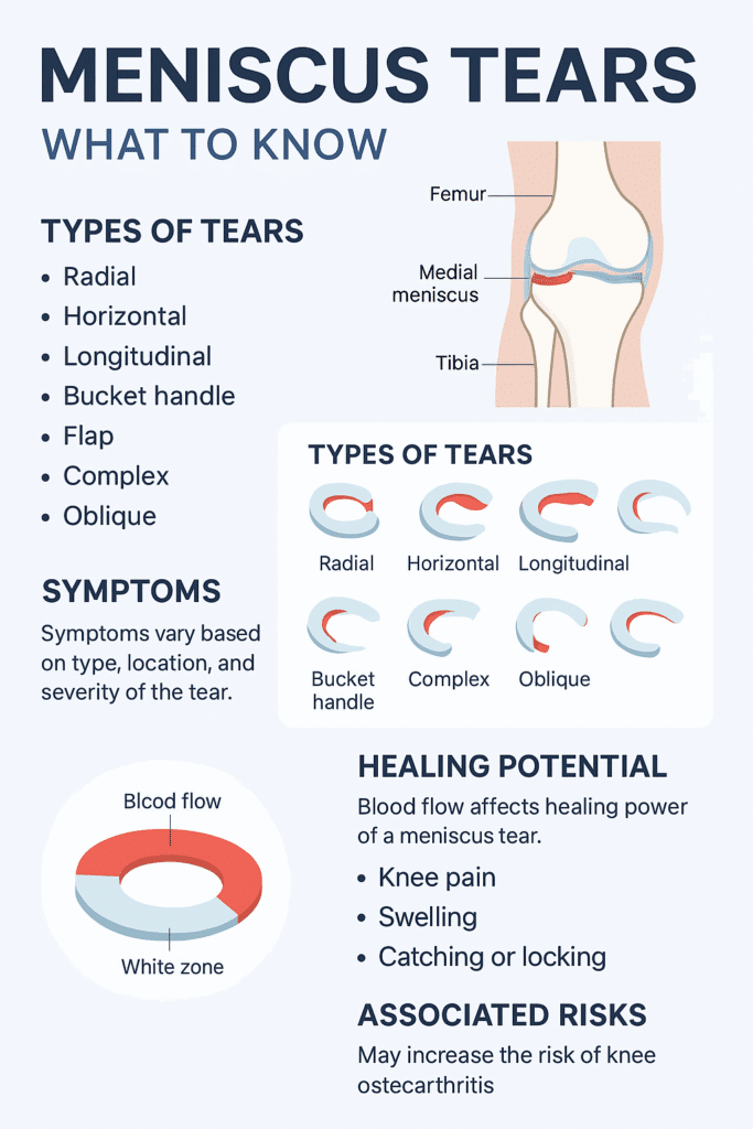
Meniscus tears are classified based on tear shape and tear location. This classification helps determine whether the injury may heal with rest and therapy or if it needs surgical treatment. If you're looking to understand the type of tear you have, we have broken them down below:
Meniscus Tear Shapes
1. Meniscus Radial Tear
A radial tear cuts straight across the meniscus from the inner edge toward the outer rim, similar to the spoke of a wheel. These are common and usually occur in areas with poor blood flow, which limits the body’s ability to heal the tear on its own. Treatment often involves trimming the damaged section.
2. Meniscus Horizontal Tear
A horizontal tear runs between the upper and lower layers of the meniscus, dividing it in half like a sandwich. These tears are more common in older adults and may be repairable if located in the outer region of the meniscus, where blood supply is better.
3. Meniscus Vertical (Longitudinal) Tear
This tear runs lengthwise along the curve of the meniscus, following its C-shape. It is often seen in younger, athletic individuals and may respond well to surgical repair, especially when located in the outer third of the meniscus.
4. Meniscus Bucket Handle Tear
A bucket handle tear is a severe form of a vertical tear. In this case, a large portion of the meniscus flips inward into the knee joint, making it difficult to bend or straighten the knee. It often causes locking and typically requires surgery to correct.
5. Meniscus Flap Tear
A flap tear results in a loose piece of cartilage that creates an uneven edge. This flap may shift with knee motion, causing clicking, catching, or locking. If symptoms are persistent, the loose section is often trimmed during a minor procedure.
6. Meniscus Complex Tear
A complex tear includes multiple tear patterns—usually both radial and horizontal—and often occurs in worn or degenerated menisci. These are difficult to repair and typically require removal of the damaged sections.
7. Meniscus Oblique (Parrot Beak) Tear
An oblique tear forms at an angle, creating a curved flap that resembles a parrot’s beak. The flap can catch in the joint and lead to sharp pain or instability. Surgical trimming is often used if the tear is unstable.
Meniscus Tear Locations
In addition to the shape of the tear, the location is a major factor in symptoms and treatment decisions. These are the meniscus tear locations:
Medial meniscus tear – inside of the knee
Lateral meniscus tear – outside of the knee
Posterior horn tear – back portion of the meniscus
Anterior horn tear – front portion of the meniscus
Meniscus root tear – where the meniscus anchors to the bone
Each location has different mechanical demands, and tears in different areas may feel different or affect how the knee moves.
Location and Healing Potential
The meniscus has three zones based on blood supply that affect how well a tear can heal:
Red zone (outer third) – good blood supply; better chance of healing on its own or after repair
White zone (inner third) – poor blood flow; limited natural healing
Red-white zone (middle third) – transitional; healing depends on tear size and stability
Long-Term Effects of Untreated Meniscus Tears
How long does it take a meniscus tear to heal? The answer is, it depends. However, if a torn meniscus is left untreated or heals poorly, it can lead to:
Chronic knee instability
Frequent swelling and inflammation
Faster joint cartilage breakdown
Higher risk of developing osteoarthritis
Early diagnosis, combined with the right treatment approach, helps protect long-term knee function and mobility.
This blog post is meant to be informative and should not act as a self-diagnosis tool. If you’d like to see one of our doctors, please contact us here.
Understanding Bowlegs: Causes, Symptoms, and Treatment Options
What Are Bowlegs?
Bowlegs, medically known as genu varum, is a condition where the legs curve outward at the knees while the ankles remain together. It is common in infants due to their fetal positioning in the womb, and in most cases, the legs straighten naturally as the child grows and begins to walk. However, if the bowing persists beyond early childhood or appears later in life, it may indicate an underlying condition requiring medical attention.
Bowlegs can affect a person’s posture and how they walk (gait), potentially leading to joint misalignment and stress over time. The degree of bowing can vary from mild to severe, and its impact on daily activities depends on a person's underlying cause and individual anatomy.
What Does It Feel Like to Have Bowlegs?
Individuals may experience these bowleg symptoms:
A noticeable outward curvature of the legs when standing with feet together
Knee and hip discomfort, especially after physical activity
Instability while walking or running
Limited mobility, particularly in the hips and knees
Increased stress on the joints, which can lead to arthritis over time
Fatigue in the legs due to inefficient movement patterns
Lower back pain resulting from compensatory postural adjustments
The severity of symptoms varies, with some individuals experiencing minimal discomfort, while others may have significant pain and difficulty moving.
What Causes Bowlegs?
Several factors and conditions can contribute to bowlegs, including:
Physiological Bowing – Common in infants and young children, this naturally corrects itself by age 3 to 4.
Blount’s Disease – A growth disorder affecting the tibia (shin bone) that worsens over time, requiring medical intervention. It is more prevalent in children who begin walking at an early age.
Rickets – A vitamin D deficiency leading to weakened bones and improper growth. This condition can be prevented with proper nutrition and sun exposure.
Bone Dysplasia – Abnormal bone development due to genetic conditions, often leading to long-term mobility challenges.
Paget’s Disease – A metabolic disorder that disrupts normal bone remodeling, leading to deformities. It typically affects adults and may require lifelong management.
Poorly Healed Fractures – If a broken leg bone heals incorrectly, it may result in permanent bowing and uneven weight distribution across the joints.
Achondroplasia – A genetic disorder that leads to dwarfism and often results in bowlegs. This condition is associated with shorter limbs and joint laxity.
Lead or Fluoride Poisoning – Exposure to high levels of these substances can interfere with normal bone development, potentially leading to long-term health issues beyond bowlegs.
Why you can trust us:
We have multiple highly specialized, board-certified, fellowship-trained orthopaedic surgeons.
We know that we serve people - actual humans - not random orthopaedic conditions. That drives us to compassionate care.
The world of orthpaedics is constantly evolving. Our orthopaedic surgeons are constantly evaluating new techniques, tools and methods to serve our community even better.
We provide outcome-focused treatment. We work with our patients to achieve their goals, all while developing custom treatment plans that fit our patient's lives.
While mild bowing in young children often corrects itself, medical evaluation is necessary if:
The bowing persists beyond age 3
One leg is more bowed than the other
The condition worsens over time
There is pain, instability, or difficulty walking
An underlying disease is suspected
The bowing is accompanied by other developmental delays or bone abnormalities
Early intervention is crucial in cases linked to nutritional deficiencies or growth disorders, as timely treatment can improve outcomes.
If you are an adult dealing with bowlegs, you should consider seeking treatment if you experience:
Pain or discomfort in the knees, hips, or ankles.
Difficulty walking or standing for long periods.
Worsening bowing or noticeable asymmetry.
Frequent joint stiffness or instability.
Early signs of arthritis, such as swelling or reduced mobility.
If bowleggedness is interfering with daily activities or causing joint damage, consult a doctor or orthopedic specialist for evaluation and treatment options like physical therapy, braces, or surgery if necessary.
Why Treat Bowlegs?
Untreated bowlegs can lead to complications, including:
Increased risk of arthritis, particularly in the knees and hips, due to uneven joint stress
Gait abnormalities affecting posture and movement efficiency
Lower back pain from altered spinal alignment
Joint instability, making physical activities more challenging
Increased risk of injury, particularly in athletes and active individuals
Muscle imbalances, as the body compensates for misalignment
Diminished quality of life, as chronic pain and mobility issues impact daily activities
Treatment Options for Bowlegs
The appropriate treatment for bowlegs is individual and depends on the severity and underlying cause of the condition.
Non-Surgical Treatments for Bowlegs
Bracing or Orthotics – Used in children to guide proper bone growth and support alignment correction.
Physical Therapy – Strengthening muscles, improving posture, and enhancing mobility to alleviate discomfort and prevent progression.
Nutritional Supplements – Correcting deficiencies in vitamin D or calcium for cases related to rickets, often combined with diet modifications.
Weight Management – Reducing excess body weight to minimize joint stress and prevent the worsening of the condition.
Low-Impact Exercises – Activities such as swimming or cycling can help strengthen leg muscles without excessive joint strain.
Surgical Treatment for Bowlegs
For severe cases, especially in teenagers and adults, surgery may be necessary. Osteotomy is the most common procedure, which involves:
Making an incision near the knee.
Cutting and realigning the tibia (or femur, in some cases) to correct the bowing.
Inserting bone grafts and securing the bone with plates, screws, or external fixators.
Undergoing physical therapy post-surgery to restore mobility and prevent stiffness.
Recovery from an osteotomy can take 3 to 6 months, it depends on the complexity of the procedure. Patients typically require crutches or braces initially and gradually return to normal activities.
What Type of Orthopedic Doctor to See?
If you suspect bowlegs in yourself or your child, consult a pediatric orthopedic specialist for children or an adult orthopedic surgeon for adults.
Bowlegs can be a normal part of early childhood development but may also indicate underlying health issues. Early diagnosis and appropriate treatment can prevent complications, improve mobility, and enhance overall quality of life. If you or a loved one has persistent bowlegs, consult one of our orthopedic specialists to explore the best treatment options. Understanding the causes and potential impact of bowlegs can empower you to seek timely medical advice and make informed decisions about their health.
This blog post is meant to be informative and should not act as a self-diagnosis tool. If you’d like to see one of our doctors, please contact us here.
Treating Runner's Knee Through Strengthening Exercises
Effective exercises for runner's knee focus on strengthening muscles around the knee, improving flexibility, and enhancing stability. These exercises reduce stress on the kneecap and improve joint alignment. Runner's knee exercises are the cornerstone of your knee rehabilitation.
Straight Leg Raises
Purpose: Strengthens the quadriceps without placing stress on the knee joint.
How-To:
Lie flat on your back with one leg bent and the other straight.
Tighten the muscles in your straight leg and lift it to the height of the bent knee.
Hold for 2–3 seconds, then slowly lower the leg. Repeat 10–15 times per leg.
Why It Works for Runner's Knee:
Strengthens the quadriceps to support the knee.
Improves joint stability without bending the knee.
Reduces stress on the patellofemoral joint
Mini Squats
Purpose: Builds strength in key knee-supporting muscles while avoiding excessive stress.
How-To:
Stand with feet shoulder-width apart, near a chair or wall for balance.
Slowly squat down to about a 45-degree angle, keeping your knees aligned over your toes.
Hold for 1–2 seconds, then return to standing. Repeat 10–15 times.
Why It Works for Runner's Knee:
Strengthens the quadriceps, hamstrings, and glutes.
Helps stabilize the knee joint during movement.
Promotes proper alignment to reduce knee strain.
Clamshells
Purpose: Strengthens hip muscles to improve knee alignment and stability.
How-To:
Lie on your side with knees bent at a 90-degree angle.
Keeping your feet together, lift the top knee as high as possible without moving your pelvis.
Lower the knee slowly. Repeat 10–15 times per side.
Why It Works for Runner's Knee:
Targets the gluteus medius for better hip stability.
Prevents inward knee movement that strains the joint.
Enhances overall leg alignment during activities.
Step-Ups
Purpose: Strengthens lower-body muscles while mimicking functional movements.
How-To:
Stand in front of a sturdy step or platform.
Step onto the platform with one foot, driving through your heel to bring the other foot up.
Step back down with the same foot and repeat. Perform 10–12 reps per leg.
Why It Works for Runner's Knee:
Builds strength in the quadriceps, glutes, and calves.
Encourages proper knee tracking over the toes.
Improves balance and stability in functional movement.
Quadriceps Stretch
Purpose: Relieves tightness in the thigh muscles to reduce stress on the knee.
How-To:
Stand on one leg, holding onto a wall or chair for support.
Pull the opposite foot toward your glutes, keeping your knees close together.
Hold for 20–30 seconds, then switch sides.
Why It Works for Runner's Knee:
Loosens tight quadriceps that can pull on the kneecap.
Improves flexibility and joint movement.
Reduces strain on the patellofemoral joint.
Hamstring Stretch
Purpose: Reduces tension in the back of the thigh to improve knee alignment.
How-To:
Sit on the floor with one leg extended and the other bent.
Reach toward the toes of the extended leg while keeping your back straight.
Hold for 20–30 seconds, then switch legs.
Why It Works for Runner's Knee:
Loosens tight hamstrings that can pull the knee out of alignment.
Enhances flexibility and joint mobility.
Helps balance muscle tension around the knee.
Calf Stretch
Purpose: Eases tension in the lower leg to reduce stress on the knee.
How-To:
Stand facing a wall, placing your hands on it for support.
Step one foot back, keeping it straight, while bending the front knee.
Press the back heel into the floor and hold for 20–30 seconds. Switch legs.
Why It Works for Runner's Knee:
Loosens tight calves to improve shock absorption.
Enhances lower leg alignment during activity.
Reduces stress transmitted to the knee joint.
Single-Leg Balance
Purpose: Enhances knee stability and improves proprioception.
How-To:
Stand on one leg with your hands on your hips or out for balance.
Hold for 20–30 seconds, gradually increasing the time as your balance improves.
For added difficulty, try closing your eyes or standing on a soft surface.
Why It Works for Runner's Knee:
Strengthens stabilizing muscles around the knee and ankle.
Improves body awareness and balance.
Reduces improper knee alignment during movement.
Side-Lying Leg Lifts
Purpose: Strengthens outer thigh muscles for improved knee tracking.
How-To:
Lie on your side with your legs straight.
Lift the top leg slowly, keeping it in line with your body.
Lower it back down without letting it rest. Repeat 10–15 times per side.
Why It Works for Runner's Knee:
Strengthens the outer thigh to stabilize the knee.
Reduces lateral instability.
Improves overall knee alignment during movement.
IT Band Foam Rolling
Purpose: Relieves tension in the iliotibial band to reduce knee strain.
How-To:
Lie on your side with a foam roller under the outer thigh.
Roll slowly from your hip to just above the knee, pausing on tight spots.
Perform for 1–2 minutes per side.
Why It Works for Runner's Knee:
Loosens the IT band, which can pull on the knee.
Improves flexibility and reduces friction around the knee joint.
Enhances mobility for smoother movement patterns.
If you feel a tight, achy feeling behind your knee, it might be a Baker's cyst.
Daily life with a Baker's cyst often feels like an uninvited house guest, quietly making its presence known with discomfort or swelling behind your knee. However, understanding Baker's cyst symptoms and causes can bring you a sense of empowerment and control, time to kick that house guest out. If you have a Baker's cyst, you may notice the area feeling tight or achy, particularly after standing or walking long. Simple activities like climbing stairs, kneeling, or even fully extending your leg might become challenging. As an orthopaedic surgeon at the forefront of musculoskeletal care, I've seen how disruptive Baker's cysts can be to daily life. It's not just the physical discomfort—there's also the lingering uncertainty about what this swelling means and how to manage it.
Why you can trust us:
We have multiple highly specialized, board-certified, fellowship-trained orthopaedic surgeons.
We know that we serve people - actual humans - not random orthopaedic conditions. That drives us to compassionate care.
The world of orthpaedics is constantly evolving. Our orthopaedic surgeons are constantly evaluating new techniques, tools and methods to serve our community even better.
We provide outcome-focused treatment. We work with our patients to achieve their goals, all while developing custom treatment plans that fit our patient's lives.
A Baker's cyst (known as a popliteal cyst) is a fluid-filled sac that forms behind your knee. It develops when excess joint fluid—known as synovial fluid—accumulates in the popliteal bursa; this small sac serves to help cushion the knee joint. This accumulation causes swelling and, in some cases, discomfort.
The Baker's cyst is usually a symptom of an underlying issue, such as arthritis or a meniscus tear. While the cyst can vary in size, it can become large enough to restrict knee movement and cause significant pain.
What are Baker's Cyst Symptoms?
Bakers cyst symptoms can vary from person to person. Many individuals experience no symptoms at all, while others may notice:
A bulging mass or lump behind the knee
Tightness or stiffness in the knee
Pain in the back of the knee, especially after standing or moving around
Swelling that can extend down the calf in some cases
Limited range of motion in the knee
A clicking or popping sensation with movement
In rare cases, the cyst can rupture, causing a sharp pain with swelling in your lower leg that mimics the symptoms of a blood clot; this can be a medical emergency and should be addressed immediately. Additional symptoms of a ruptured Baker's cyst may include severe pain, sudden swelling, and difficulty bearing weight on the affected leg.
Where Do Bakers Cysts Form, and Why?
Baker's cysts form in the popliteal space at the back of the knee joint. This space contains the popliteal bursa, which is prone to filling with excess synovial fluid under certain conditions. When the knee experiences inflammation due to arthritis, injury, or other joint issues, the body naturally produces more fluid to cushion the joint. However, when there is too much fluid, it can leak into the bursa and form a cyst.
The location behind the knee makes this area particularly vulnerable to fluid buildup because of its anatomy and function in movement, especially when the knee joint is stressed or overused.
What Causes a Baker's Cyst?
Several underlying conditions can lead to the formation of a Baker's cyst:
Osteoarthritis: As the most common cause, this wear-and-tear condition inflames the knee joint, increasing fluid production.
Rheumatoid arthritis: Inflammatory arthritis can cause excessive fluid to accumulate in the affected joint.
Meniscus tear: This injury is a tear in the cartilage of your knee and can trigger inflammation, leading to cyst development.
Knee injuries: Trauma or repetitive stress can inflame the knee joint, prompting fluid buildup.
Gout or other joint issues: Conditions that cause swelling in the knee may result in forming a Baker's cyst.
Is There a Way to Prevent a Baker's Cyst?
While there's no guaranteed way to prevent a Baker's cyst, addressing the underlying causes can reduce the risk of development.
Proper management of arthritis
Regular low-impact exercises
Maintaining a healthy weight to reduce the stress imposed on your knees
Protecting your knees from injury with proper gear and technique during physical activities can also help minimize the chances of developing a cyst.
How to Care for a Baker's Cyst Once You Have One
Once you've been diagnosed with a Baker's cyst, there are several steps you can take to manage the condition and reduce discomfort:
At-Home Treatments
Rest: Limiting activities that aggravate the knee joint can help reduce swelling.
Ice: Applying ice to the back of the knee for 15-20 minutes can help relieve pain and reduce swelling.
Compression: Using a compression bandage can help manage swelling by limiting fluid buildup in the area.
Elevation: Keeping your leg elevated when resting can assist in reducing fluid accumulation and discomfort.
Over-the-counter pain relief: Nonsteroidal anti-inflammatory drugs (NSAIDs) like ibuprofen can help manage pain and inflammation.
What Not to Do When You Have a Baker's Cyst
What not to do when you have a Baker's cyst is just as important as understanding what you should do.
Avoid if You Have a Baker's Cyst
Excessive activity: Overusing the knee through high-impact exercises can worsen the condition.
Ignoring symptoms: Don't wait for the pain or swelling to go away on its own—delaying care can worsen the condition.
Assuming it's just a muscle strain: Misdiagnosing a Baker's cyst as a simple muscle strain can lead to improper treatment and an increased risk of rupture.
When to Seek Help from POA's Orthopaedic Surgeons
If your Baker's cyst persists despite home treatment, causes significant discomfort, or interferes with your daily activities, it's time to consult an orthopaedic surgeon. Seeking professional care can provide reassurance and confidence in your decision-making. At POA, our team of expert orthopaedic surgeons specializes in diagnosing and treating conditions like Baker's cysts and any potential underlying causes like arthritis or meniscus tears.
What Type of Orthopaedic Specialist Should You See?
If you are looking for a doctor to help with a Baker's cyst, you'll want to see an orthopaedic surgeon specializing in knee conditions. At POA, our knee specialists are highly experienced in managing the symptoms and root causes of Baker's cysts, ensuring you receive personalized, comprehensive care.
Meet Our Orthopaedic Knee Specialists
What Does Treatment Look Like from a Medical Standpoint?
Treatment for a Baker's cyst often begins conservatively. In many cases, if the underlying condition causing the cyst is treated, the cyst itself will improve. Here's what medical treatment might involve:
Aspiration: For aspiration of the cyst, your orthopaedic surgeon uses a needle to drain the excess fluid from the cyst, which can provide temporary relief from swelling and discomfort.
Corticosteroid Injections: These anti-inflammatory injections can help reduce swelling and manage pain associated with the cyst.
Physical Therapy: Targeted exercises can help improve joint function, reduce fluid buildup, and manage pain.
Surgery: In cases where the cyst is large, recurrent, or caused by an underlying structural issue, surgery may be necessary to remove the cyst or repair the damaged joint tissue.
Whether you need imaging, joint aspiration, or surgical intervention, our surgeons have the expertise to guide you through the best treatment options.
When to Seek Urgent Care
While Baker's cysts are rarely life-threatening, there are certain instances where we recommend seeking urgent care. If your cyst ruptures, you may experience severe pain, swelling, and bruising in your lower leg; this can be mistaken for a blood clot. If you experience this, immediately seeking medical attention is crucial to rule out the possibility of a more serious condition such as a deep vein thrombosis (DVT).
Need Care Now? POA Has Six Urgent Care Facilities
Monroe
11 Centre Drive Monroe Twp., NJ 08831
Plainsboro
5 Plainsboro Road, Suite 100 Plainsboro, NJ 08536
Robbinsville
1 Union Street Suite 305 Robbinsville, NJ 08691
Princeton
325 Princeton Avenue Princeton, NJ 08540
Hillsborough
315 US Highway 206 Hillsborough Township, NJ 08844
Living with a Baker's cyst can be frustrating, but with the proper care and treatment, it can be entirely manageable. Understanding the condition, starting to address the root causes, and seeking the expertise of an orthopaedic surgeon can make a world of difference.
If you're experiencing Baker's cyst symptoms, don't let the pain or swelling limit your daily life. The knee specialists at POA are here to provide comprehensive, personalized care and guide you through every step of your treatment plan.
Schedule an appointment today with one of POA's expert orthopaedic surgeons and take the first step toward relief!
Does a Baker's Cyst Have Anything to do With Being a Baker?
A Baker’s cyst is not named after bakers or anything related to baking! Instead, it’s named after the British surgeon William Morrant Baker, who first described the condition in the late 1800s. The cyst itself is a fluid-filled sac that forms behind the knee, often due to issues like arthritis or a meniscus tear. While it might sound like something that might happen to a baker, the name is purely a nod to the doctor’s contributions to understanding this knee issue. So, if you ever hear someone mention a Baker’s cyst, remember it's all about medical history and not about pastry!
This blog post is meant to be informative and should not act as a self-diagnosis tool. If you’d like to see one of our doctors, please contact us here.
MCL Tear: Symptoms, Treatment Options, and Recovery
Living with an MCL tear presents a series of daily challenges that can disrupt your routine and overall well-being. From the moment you wake up, simple activities like getting out of bed or walking to the kitchen require extra care and patience due to instability in your knee. Mobility is limited, making everyday tasks such as climbing stairs or carrying groceries a bit more demanding. However, as you see care, adopt new strategies, and build resilience, with patience, determination, and a strong support system, you can navigate the journey of recovery and emerge stronger.
What is an MCL Tear?
A medial collateral ligament (MCL) tear is an injury to one of the major ligaments in the knee. The MCL on the knee's inner side helps stabilize the joint and prevents the knee from bending inward. MCL tears commonly occur in sports that involve sudden changes in direction, twisting, or direct blows to the knee, such as football, soccer, and skiing
Symptoms of an MCL Tear
The symptoms of an MCL tear can vary depending on the severity of the injury but typically include:
Pain on the inner side of the knee
Swelling around the knee joint
Instability or a feeling that the knee may "give out."
Difficulty walking or bearing weight on the affected leg
Stiffness and reduced range of motion in the knee
These symptoms can range from mild discomfort to severe pain, significantly affecting your ability to perform everyday activities and participate in sports.
Treatment Options for an MCL Tear
Treatment for an MCL tear depends on the severity of the injury. Here are the main treatment options:
1. Rest and Ice
Rest and ice can help reduce pain and swelling for mild MCL tears. Avoid activities that put stress on the knee, and apply ice to the affected area for 20 minutes several times daily.
2. Compression and Elevation
A compression bandage can help control swelling while elevating the leg above heart level, reducing inflammation and promoting healing.
3. Physical Therapy
Physical therapy is crucial for recovering from an MCL tear. A physical therapist can design a rehabilitation program that includes:
Range-of-motion exercises to prevent stiffness
Strengthening exercises for the muscles around the knee
Balance and stability exercises to improve knee function
Gradual return-to-sport activities
4. Medications
Over-the-counter pain relievers, such as ibuprofen or acetaminophen, can help manage pain and reduce inflammation. In some cases, your doctor may prescribe stronger medications for short-term pain relief.
5. Bracing
Wearing a knee brace can provide additional support and stability, especially during the initial stages of recovery. By limiting excessive knee movement, the brace helps prevent further injury.
6. Surgery
Surgery is usually reserved for severe MCL tears or when other structures in the knee, such as the anterior cruciate ligament (ACL), are also damaged. Surgical options include:
Repairing the torn ligament with sutures
Reconstructing the ligament using a graft from another part of the body
When to See an Orthopaedic Specialist
You should see an orthopaedic specialist if:
Your knee pain and swelling do not improve with rest and self-care
You experience significant instability or difficulty bearing weight on the affected leg
You have concerns about returning to sports or physical activities
An orthopaedic specialist can assess the severity of your injury, perform imaging tests, and develop a tailored treatment plan to ensure optimal recovery.
Why you can trust us:
We have multiple Orthopaedic Spine & Back Specialists who treat spine & back-related injuries every day.
We have the Spine and Back Institute with a team of world-recognized back specialists, orthopaedists, and therapists who have focused solely on back, neck, and spine health issues for over 15 years.
We have a whole-body health approach when it comes to orthopaedic spine, neck, and back health. The health of the entire body is connected to your back.
MCL tears can significantly impact your mobility and quality of life. Early intervention and proper treatment are essential for a successful recovery. If you suspect you have an MCL tear or are experiencing persistent knee pain, schedule an appointment with an orthopaedic specialist to receive a comprehensive evaluation and personalized treatment plan.
By taking action, you can prevent further damage, reduce pain, and return to your normal activities more quickly. Don't let an MCL tear keep you on the sidelines – seek professional help and start your path to recovery today.
This blog post is meant to be informative and should not act as a self-diagnosis tool. If you’d like to see one of our doctors, please contact us here.
Understanding Bone Spurs
Have you ever experienced a sharp pain in your foot when taking your first steps in the morning or a nagging ache in your shoulder that won't go away? You might be dealing with a foot or shoulder bone spur. These small, bony growths can develop in various parts of the body, causing discomfort and limiting your mobility.
What is a Bone Spur?
A bone spur, also known as osteophyte, is a bony projection that forms along the edges of bones. These growths typically develop where bones meet each other in the joints. While bone spurs are not necessarily painful, they can cause problems when they rub against nearby nerves or tissues.
What Causes Bone Spurs?
Bone spurs often develop in response to pressure, rubbing, or stress on a bone over time.
Common causes include:
Osteoarthritis
Osteoarthritis is a degenerative joint disease and one of the most common causes of bone spurs. As the protective cartilage between bones wears down over time, the body may respond by forming extra bone around the affected joint edges, resulting in bone spurs.
Repetitive Stress or Overuse
Activities that involve repetitive motions or stress on specific joints, such as regularly lifting heavy objects, running, or jumping, can lead to the formation of bone spurs. Over time, the constant pressure on the bones can cause them to develop extra bony growths.
Age-related Wear and Tear
As people age, the cartilage in their joints naturally begins to deteriorate. This can result in increased bone friction, leading to bone spurs, especially in weight-bearing joints like the spine, knees, or hips.
Joint Diseases
Inflammatory joint conditions such as rheumatoid arthritis, ankylosing spondylitis, or gout can cause inflammation and damage to the joint tissues, leading to bone spur formation as the body tries to repair itself.
Poor Footwear
Regularly wearing footwear that doesn't provide adequate support or has an improper fit, such as narrow shoes and high heels, can lead to the development of bone spurs in the feet, particularly in the heel area.
Trauma or Injury
Previous joint injuries, such as fractures, dislocations, or ligament tears, can cause the body to produce extra bone in the healing process; this has the potential to lead to the formation of bone spurs in the affected area.
Genetics
Some individuals can have a genetic predisposition to developing bone spurs. Certain inherited conditions or structural abnormalities can increase the likelihood of spur formation, even without other contributing factors.
Obesity
Excess body weight is known to put added stress on joints, such as the spine, knees, and hips. Over time, this increased pressure can lead to wear and tear and the development of bone spurs.
Understanding these common causes can help individuals take preventive measures and seek appropriate treatment if they experience symptoms of bone spurs.
Why you can trust us:
We have multiple specialists who treat these conditions every day.
Our orthopaedic doctors are specialized, which means you can see a doctor who works solely with the part of the body you are having issues with.
We have a whole-body health approach when it comes to orthopaedic health, and along with orthopaedic specialists, we have a team of physiatrists and physical therapists here to help you get back to the things you love.
A bone spur can form in various parts of the body, including:
Foot:
A bone spur in the foot, especially the heel (heel spurs), can cause sharp pain, particularly during activities like walking or standing.
Symptoms: Heel pain, tenderness, difficulty bearing weight on the affected foot.
Shoulder:
A shoulder bone spur can form on the acromion (the bony process on the shoulder blade) or on the joint surfaces where the collarbone and shoulder blade meet. These spurs can result from overuse, injury, or age-related wear and tear.
Symptoms: Shoulder pain, stiffness, weakness, difficulty raising the arm.
Elbows:
Bone spurs in the elbows can develop in conditions like tennis elbow (lateral epicondylitis) or golfer's elbow (medial epicondylitis), where the tendons attaching to the elbow become inflamed and may develop spurs over time.
Symptoms: Elbow pain, tenderness, difficulty bending or straightening the arm.
Hands:
Bone spurs can form in the joints of the fingers or thumbs, often due to osteoarthritis or repetitive use of the hands.
Symptoms:
Joint pain,
swelling,
stiffness,
bony nodules.
Knees:
Bone spurs in the knees can form around the joint due to osteoarthritis or other conditions that cause wear and tear on the joint cartilage.
Symptoms: Knee pain, swelling, stiffness, decreased range of motion.
Spine:
Bone spurs in the spine, also called osteophytes, can occur along the edges of vertebrae. They can develop due to degenerative conditions where the cartilage between vertebrae breaks down, like osteoarthritis, causing bone-on-bone contact.
Symptoms: Back pain, stiffness, radiating pain or numbness.
Hips:
Hip bone spurs can develop in the hip joint, particularly in individuals with hip osteoarthritis or a condition known as femoroacetabular impingement (FAI), where abnormal contact between the ball and socket of the hip joint occurs.
Symptoms:
Hip pain,
stiffness,
reduced range of motion.
What Does a Bone Spur Feel Like?
While your symptoms can vary depending on the location of the bone spur, common signs include the following:
If you suspect you have a bone spur, it's essential to consult with a healthcare professional. Your doctor, typically an orthopedic surgeon, will perform a physical examination, ask questions, and may order diagnostic tests such as X-rays or MRI scans to confirm the diagnosis.
Bone Spur Treatment Options
Fortunately, there are several treatment options available for bone spurs, ranging from conservative measures to surgical intervention:
Medication: Over-the-counter pain relievers may be recommended to help alleviate discomfort. OTC pain relievers such as ibuprofen or acetaminophen.
Physical Therapy: Stretching and strengthening exercises are often recommended because they can improve flexibility and reduce symptoms.
Orthotics: With a foot bone spur, orthotics may be recommended. These custom shoe inserts help provide support and relieve pressure on the affected area.
Injections: Corticosteroid injections may be recommended to reduce the inflammation and help alleviate your pain.
Surgery: In more severe cases where conservative treatments are unsuccessful, it may be recommended to have surgical removal of the bone spur.
How to Dissolve Bone Spurs Naturally
While there's limited scientific evidence to support natural remedies for dissolving bone spurs, some people find relief through:
A healthy diet consisting of anti-inflammatory foods such as fruits, vegetables, as well as omega-3 fatty acids may help reduce inflammation.
Maintaining a healthy weight to lessen the added strain on your joints, potentially slowing the progression of bone spurs.
Some individuals report benefits from supplements like glucosamine and chondroitin, though results vary.
While there is limited scientific evidence that these techniques will dissolve bone spurs naturally, they are great lifestyle choices and will contribute to your overall well-being even if they don't.
Don't Live with Bone Spurs - Seek Help
Living with bone spurs can significantly impact your quality of life, but you don't have to suffer in silence. If you're experiencing persistent pain or discomfort, schedule an appointment with one of our specialists. They can thoroughly evaluate and recommend the most effective treatment plan to help you get back to doing what you love. Don't let bone spurs hold you back any longer!
Sequence rules uses cookies to track the order of requests a user has made and the time between requests and makes them available via Cloudflare Rules. This allows you to write rules that match valid or invalid sequences. The specific cookies used to validate sequences are called sequence cookies.
session
cf_ob_info
The cf_ob_info cookie provides information on: The HTTP Status Code returned by the origin web server. The Ray ID of the original failed request. The data center serving the traffic
session
cf_use_ob
The cf_use_ob cookie informs Cloudflare to fetch the requested resource from the Always Online cache on the designated port. Applicable values are: 0, 80, and 443. The cf_ob_info and cf_use_ob cookies are persistent cookies that expire after 30 seconds.
session
__cfwaitingroom
The __cfwaitingroom cookie is only used to track visitors that access a waiting room enabled host and path combination for a zone. Visitors using a browser that does not accept cookies cannot visit the host and path combination while the waiting room is active.
session
cf_chl_rc_i
These cookies are for internal use which allows Cloudflare to identify production issues on clients.
session
cf_chl_rc_ni
These cookies are for internal use which allows Cloudflare to identify production issues on clients.
session
cf_chl_rc_m
These cookies are for internal use which allows Cloudflare to identify production issues on clients.
session
__cfruid
Used by the content network, Cloudflare, to identify trusted web traffic.
session
__cf_bm
Cloudflare's bot products identify and mitigate automated traffic to protect your site from bad bots. Cloudflare places the __cf_bm cookie on End User devices that access Customer sites that are protected by Bot Management or Bot Fight Mode. The __cf_bm cookie is necessary for the proper functioning of these bot solutions.
session
__cflb
When enabling session affinity with Cloudflare Load Balancer, Cloudflare sets a __cflb cookie with a unique value on the first response to the requesting client. Cloudflare routes future requests to the same origin, optimizing network resource usage. In the event of a failover, Cloudflare sets a new __cflb cookie to direct future requests to the failover pool.
session
_cfuvid
The _cfuvid cookie is only set when a site uses this option in a Rate Limiting Rule, and is only used to allow the Cloudflare WAF to distinguish individual users who share the same IP address.
session
cf_clearance
Whether a CAPTCHA or Javascript challenge has been solved.
session
Google reCAPTCHA helps protect websites from spam and abuse by verifying user interactions through challenges.
Name
Description
Duration
_GRECAPTCHA
Google reCAPTCHA sets a necessary cookie (_GRECAPTCHA) when executed for the purpose of providing its risk analysis.
179 days
Google Tag Manager simplifies the management of marketing tags on your website without code changes.
Name
Description
Duration
cookiePreferences
Registers cookie preferences of a user
2 years
td
Registers statistical data on users' behaviour on the website. Used for internal analytics by the website operator.
session
These cookies are used for managing login functionality on this website.
Name
Description
Duration
wordpress_logged_in
Used to store logged-in users.
Persistent
wordpress_sec
Used to track the user across multiple sessions.
15 days
wordpress_test_cookie
Used to determine if cookies are enabled.
Session
WPForms is a user-friendly WordPress plugin for creating custom forms with drag-and-drop functionality.
Name
Description
Duration
wpfuuid
Used to track user interactions with forms.
11 years
Statistics cookies collect information anonymously. This information helps us understand how visitors use our website.
Google Analytics is a powerful tool that tracks and analyzes website traffic for informed marketing decisions.
Used to distinguish new sessions and visits. This cookie is set when the GA.js javascript library is loaded and there is no existing __utmb cookie. The cookie is updated every time data is sent to the Google Analytics server.
30 minutes after last activity
__utmc
Used only with old Urchin versions of Google Analytics and not with GA.js. Was used to distinguish between new sessions and visits at the end of a session.
End of session (browser)
__utmz
Contains information about the traffic source or campaign that directed user to the website. The cookie is set when the GA.js javascript is loaded and updated when data is sent to the Google Anaytics server
6 months after last activity
__utmv
Contains custom information set by the web developer via the _setCustomVar method in Google Analytics. This cookie is updated every time new data is sent to the Google Analytics server.
2 years after last activity
__utmx
Used to determine whether a user is included in an A / B or Multivariate test.
18 months
_ga
ID used to identify users
2 years
_gali
Used by Google Analytics to determine which links on a page are being clicked
30 seconds
_ga_
ID used to identify users
2 years
_gid
ID used to identify users for 24 hours after last activity
24 hours
_gat
Used to monitor number of Google Analytics server requests when using Google Tag Manager
1 minute
_gac_
Contains information related to marketing campaigns of the user. These are shared with Google AdWords / Google Ads when the Google Ads and Google Analytics accounts are linked together.
90 days
__utma
ID used to identify users and sessions
2 years after last activity
__utmt
Used to monitor number of Google Analytics server requests
10 minutes
These cookies help us measure the effectiveness of our website content and understand how visitors find and use our site. This allows us to improve the experience and provide more helpful resources.
Facebook Pixel is a web analytics service that tracks and reports website traffic.