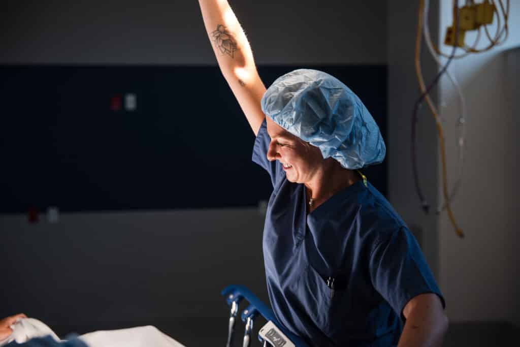
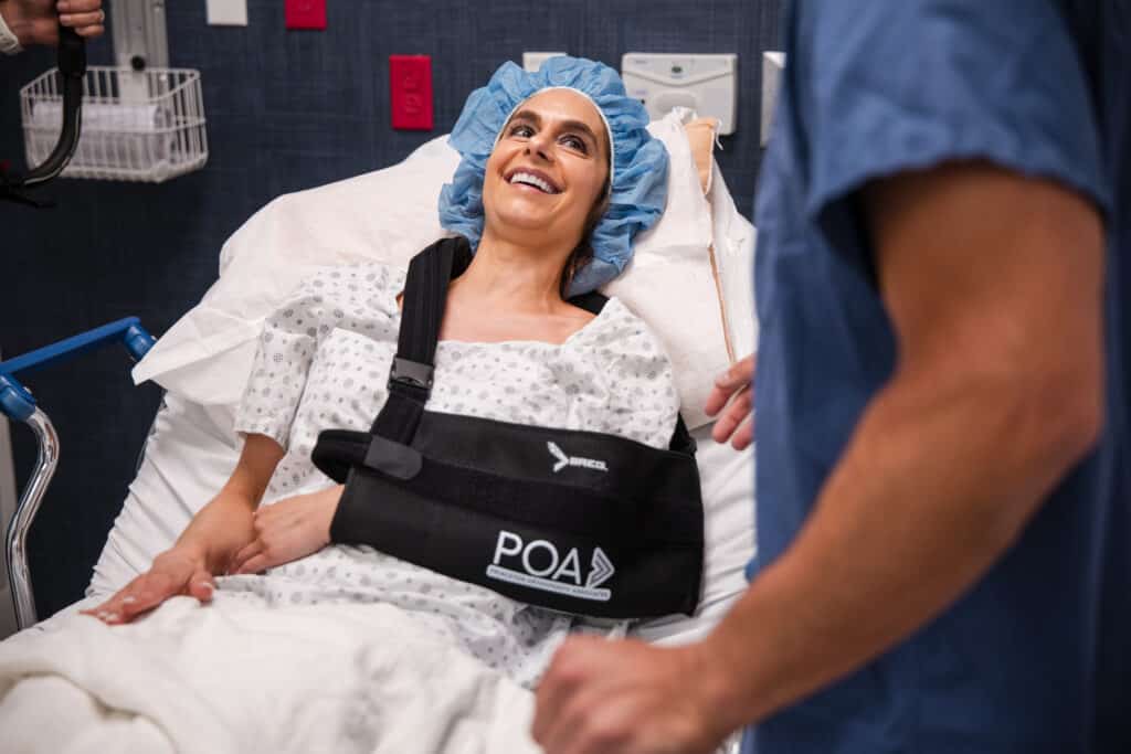
The new POA Surgery Center is a standalone surgical facility that provides patients with an exceptional surgical experience. Surgery can feel like a big deal. Because of that, our team designed a surgical fascility to help patients recover safely and comfortably, often going home the same day, with a hand-selected orthopedic team. It's the same equipment, treatments and care our surgeons would choose for their own families.
Our team made every decision about the Surgery Center with one question in mind: what is best for the patient?
Ultimately, it matters because our patients matter. With a surgery center, our team maintains control over your care. In a large hospital, orthopaedic patients share operating rooms, staff and schedules with every other specialty. At our center, every room, every team member and every piece of equipment exists for the types of surgery we perform daily.
"What we built with our new surgical center is a facility where we can provide optimal care under our control to our patients."
Why It Matters.
It was designed by our team to help patients avoid the common stress and hassle of surgery. They ge to recover where they are most comfortable: at home, cared for by people they know. Every decision about the center was made by your surgeons with care, convenience and recovery in mind.
Benefits of Surgery at the POA Surgery Center
You heal at home
At POA's surgical center, you often get to recover in your own bed and return to your life sooner than you expected.
A center just for orthopaedic care
Our surgical center is used for orthopaedic surgeries. Nothing else. That means a cleaner, less stressful environment.
Simpler, less stressful care
Parking is at the door. You are greeted by people who know who you are and why you are here, and the staff are in touch from the minute your procedure is scheduled.
A team that does orthopaedics every day
Nurses, technicians and surgeons alike are experts. We do this every day, and this is our home court.
Your surgeon makes the decisions
Your surgeon does what is best for you. Decisions are not made by an administrator or a financial analyst.
State of the art, chosen by surgeons
Our surgeons choose the operating rooms, equipment and implants they would choose for their own families.
No hospital visit
Avoid the lines. Avoid admission. Avoid the crowds. Avoid the hospital altogether.
Close to home
You do not have to travel out of state for advanced orthopaedic surgical care.
This blog post is meant to be informative and should not act as a self-diagnosis tool. If you’d like to see one of our doctors, please contact us here.
Common Knee Injuries and What You Should Know
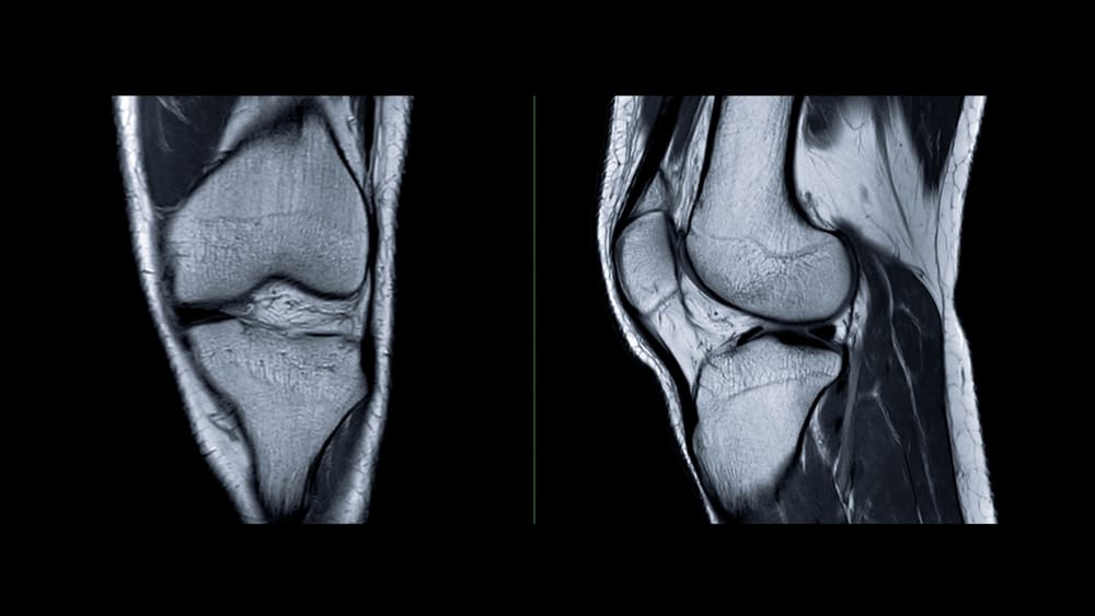
Your knees handle a lot of force each day, from walking and stairs to sports and getting up from a chair. Because the knee has many moving parts and must stay stable, injuries can range from mild sprains to problems that make it hard to move normally.
Some knee problems show up right after a twist, fall, or awkward landing. Others develop slowly from overuse. When you understand common symptoms, you can recognize concerns early and know when it is time to get medical care.
Knee Injuries Patients Often Ask About
What You Should Know
Common significant knee injuries people often ask about include ACL tears, meniscus tears, MCL injuries, PCL injuries, and patellar tendon tears.
Sports can cause these injuries, but everyday slips, sudden turns, and falls can also lead to them.
Typical symptoms include pain, swelling, a sense that the knee is not stable, locking or stiffness, and trouble bearing weight.
Not every knee injury needs surgery, but getting checked matters because some injuries can worsen if they are ignored.
Treatment usually depends on what structure is hurt, how severe it is, and your activity goals.
Early care may include rest, bracing, physical therapy, and changing what activities you do for a while.
X-rays or MRI may be used to confirm the diagnosis and check for injuries to bones, ligaments, cartilage, or tendons.
Seek orthopaedic care if the knee gives out, swells quickly, or cannot bend or straighten fully.
Why Knee Injuries Are So Common
The knee is a complex joint made up of bones, ligaments, cartilage, tendons, and muscles. It needs strong support to carry your body weight and also needs smooth motion for bending, straightening, and turning.
This mix of stability and movement makes the knee vulnerable. A forceful twist, a direct hit, a sudden stop, or an awkward landing can overload one part of the joint and lead to injury.
A Quick Comparison of Common Knee Injuries
Injury
What It Affects
Common Symptoms
ACL tear
Main stabilizing ligament in the center of the knee
Pain, swelling, feeling of looseness, discomfort with weight-bearing
Patellar tendon tear
Tendon connecting the kneecap to the shinbone
Pain below the kneecap, weakness, trouble straightening the knee
1. ACL Tears
The anterior cruciate ligament, or ACL, helps keep the knee stable, especially during cutting, pivoting, and sudden changes in direction. ACL tears are well known in sports, but they can also happen after a fall or an awkward twist.
Many people describe feeling or hearing a pop at the time of injury. Swelling often starts soon after, and the knee may feel unstable, like it could give way when you turn or try to change direction.
Often caused by pivoting, landing, or sudden stopping
May include a pop at the moment of injury
Often followed by swelling and instability
2. Meniscus Tears
The meniscus is cartilage that acts like a cushion between the thighbone and shinbone. Each knee has two menisci, and they help with shock absorption and joint stability.
A meniscus tear can happen after a twist or deep squat, especially when the foot stays planted. In some people, the tissue can also wear down with age. Symptoms may include pain, swelling, stiffness, and a feeling that the knee catches or locks.
Can happen with twisting or deep bending
May cause catching, locking, or clicking
Sometimes occurs from wear over time rather than a single injury
3. MCL Injuries
The medial collateral ligament, or MCL, runs along the inner side of the knee. It helps resist forces that push the knee inward.
MCL injuries often happen after a blow to the outside of the knee, which is common in contact sports. You may notice pain and tenderness on the inner side of the knee, along with swelling or a feeling that the knee is less stable than usual.
4. PCL Injuries
The posterior cruciate ligament, or PCL, sits behind the ACL and helps keep the shinbone from moving too far backward. PCL injuries are less common than ACL tears, but they can still cause significant symptoms.
These injuries often happen when the front of the bent knee strikes something hard, such as during a fall or motor vehicle collision. Pain, swelling, and a sense that the knee is not moving normally are common complaints.
Signs Your Knee Injury Shouldn’t Be Ignored
Swelling that starts soon after the injury
A knee that buckles or feels unstable
Locking or inability to fully bend or straighten the knee
Difficulty walking or putting weight on the leg
Pain that does not improve with rest
If you have a traumatic knee injury and you cannot bear weight, cannot actively straighten the knee, have rapid or severe swelling, see deformity, feel numbness, notice the leg is cold, or suspect dislocation or fracture, get urgent medical evaluation.
5. Patellar Tendon Tears
The patellar tendon connects your kneecap to your shinbone and helps you straighten your knee. This tendon is important for walking, climbing stairs, standing up, and jumping.
A tear can happen when the tendon is overloaded, often during a forceful jump or landing. Pain is usually felt below the kneecap, and a more serious tear can make it very hard to straighten the leg or support your weight.
How Knee Injuries Are Diagnosed
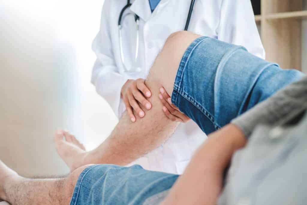
A clinician will start with your history and a physical exam. How the injury happened, where the pain is located, whether the knee swelled right away, and whether the knee feels unstable can all help guide next steps.
Imaging may also be used. X-rays can help rule out fractures, while MRI is often used to look at soft tissues like ligaments, cartilage, and tendons.
Treatment Depends on the Injury
There is no single plan for every knee injury. Care is based on which structure is hurt, how severe the damage is, your symptoms, and what you want to return to in daily life or sports.
Rest and activity changes may help protect the knee early on
Bracing can help support the knee for some ligament injuries
Physical therapy can work on strength, motion, and stability
Some problems may need surgery, especially if there is a suspected complete tendon rupture, major ligament injury, a fracture, or a locked knee
When to See an Orthopaedic Specialist
Get checked soon if your knee pain is severe, if you heard or felt a pop, or if the joint swells quickly after an injury. It also matters if the knee feels like it will give out, gets stuck, or keeps you from walking normally.
A timely evaluation can help prevent additional damage and may help you start the right treatment sooner. If the cause of your symptoms is not clear, a specialist can help you sort out what might be injured and what to do next.
Symptom
Why It Matters
Rapid swelling
Can suggest a significant ligament or internal knee injury
Instability or buckling
May point to ligament damage
Locking or catching
Can happen with meniscus tears or other internal issues
Inability to straighten the knee
May indicate a tendon problem or mechanical block
Pain with weight-bearing
Needs evaluation if it does not quickly improve
Getting Back to Daily Life Safely
A knee injury affects more than sports. It can change work tasks, sleep, exercise routines, and confidence walking on uneven ground or using stairs.
With an accurate diagnosis and a treatment plan that fits your needs, many people can return to their usual activities safely. Focus on healing and guidance, rather than pushing through pain.
This blog post is meant to be informative and should not act as a self-diagnosis tool. If you’d like to see one of our doctors, please contact us here.
Why Is My Thumb Twitching?
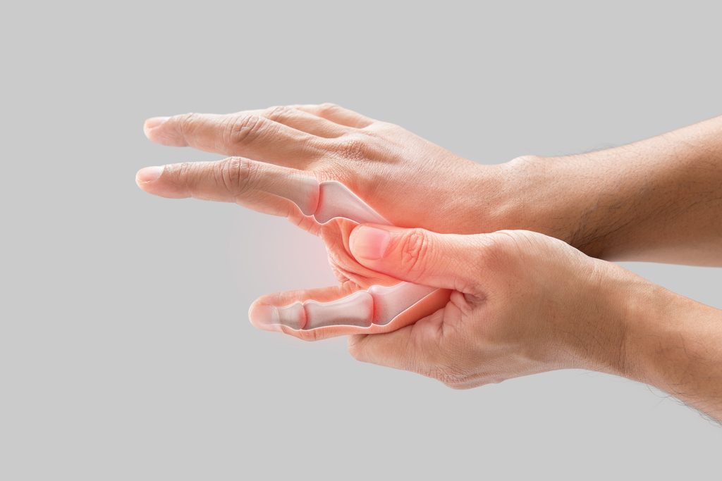
Thumb twitching can feel worrying, especially when it starts without an obvious reason. In many cases, the jerks are linked to muscle fatigue, stress, caffeine, dehydration, or insufficient sleep. Repetitive hand use can also play a role. Often, the twitching is brief and settles on its own, but it's wise to get checked if it keeps going.
Your thumb does a lot more than most people realize. It helps with texting, typing, gripping tools, cooking, and lifting. The small muscles and the tendons around your thumb work together many times a day. When they are overused, they can become irritated or fatigued, and you may notice small, on and off twitching.
Learning the common triggers can help you try safe, simple changes first. It can also guide you on when it is time to ask for an evaluation from an orthopaedic or hand specialist, especially if symptoms affect comfort or hand function.
Key Points About Thumb Twitching
What You Should Know
Thumb twitching is often related to muscle fatigue from repetitive use, stress, or too much caffeine.
Daily triggers can include typing, gaming, texting, writing, and repeated gripping.
Twitching can happen when small hand muscles get tired or irritated.
Dehydration, lack of sleep, and long periods without recovery can make twitching more likely.
Many episodes are temporary, but ongoing symptoms should not be ignored.
Twitching can also come with pain, cramping, weakness, numbness, or a tired feeling.
Nerve signals in the hand, wrist, arm, or neck can contribute to twitching.
Rest, hydration, reducing repetitive activity, and limiting stimulants may help.
If twitching lasts, worsens, or affects hand use, a medical evaluation is important.
An orthopaedic specialist can help sort out whether muscles, tendons, or nerves are involved.
What Causes a Thumb to Twitch?
A twitch is a small, involuntary change in muscle activity. In the thumb, it often comes from brief, small muscle contractions or from nerve signals that affect those muscles. This can feel like brief jumping, pulsing, or a fluttering sensation that comes and goes. The tendon or joint irritation may be present too, especially when use has been heavy.
In many people, the cause is not serious and does not last long. Overuse is a common reason, especially after lots of phone time, keyboard time, or repetitive hand work. When the hand is working in the same way for long periods, the muscles can become tired and more reactive.
Stress, caffeine, dehydration, poor sleep, and general fatigue can also make twitching more noticeable. If you notice the twitching after these triggers, it can point to a temporary muscle or nerve irritation pattern.
Common Triggers You Might Not Realize
Thumb twitching often shows up with everyday habits. Because the thumb helps with many small actions, routine tasks can place extra strain on nearby muscles and on the tendon area. The twitching may increase after a long workday or after you change your usual routine for a few days.
Frequent texting or scrolling on a smartphone
Long hours of typing or using a mouse
Video gaming or repetitive controller use
Writing, crafting, or needlework
Using hand tools or gripping repeatedly at work
Stress and muscle tension
Too much caffeine or other stimulants
Not drinking enough fluids or having poor sleep
When Overuse Is the Likely Cause
Thumb twitching that starts after a busy day of hand activity may be due to overuse. The thumb uses small muscles in the hand and forearm that can become irritated with repeated gripping, pinching, or fast finger movements. The twitching may show up more after work, at night, or after long stretches of screen time.
You might also notice the thumb or hand feels sore, tight, or tired. These feelings can happen at the same time as twitching because tendon and joint irritation can coexist with muscle fatigue. When overuse is the main issue, symptoms often improve after a short period of rest and activity changes.
Taking breaks, changing how you hold the hand, and limiting repetitive activity for several days may reduce symptoms. Gentle movement and avoiding long, steady positions can also help your muscles calm down.
Could It Be Related to a Nerve?
Sometimes thumb twitching is more than muscle fatigue. It can also happen when a nerve that helps control thumb muscles becomes irritated or compressed. This can occur in the hand, wrist, elbow, or even in the neck.
If a nerve is involved, twitching may come with other symptoms like tingling, numbness, burning pain, weakness, or trouble gripping. When twitching is paired with these signs, it is a good idea to seek medical care rather than waiting it out.
Other Symptoms That May Happen With Thumb Twitching
A twitch by itself is often less concerning than twitching plus other changes. It helps to notice what else you feel and whether the symptoms interfere with daily tasks like opening jars, holding a phone, or using tools. Keep track of when symptoms happen and what you were doing right before they started.
Thumb or hand pain
Cramping or stiffness
Numbness or tingling
Weak grip or dropping objects
Swelling near the thumb or wrist
Symptoms that spread into the fingers, forearm, or arm
Possible Causes and What They May Feel Like
Possible Cause
What You May Notice
Common Trigger
Muscle fatigue
Brief twitching or fluttering without major pain
Heavy hand use or repetitive motion
Stress or caffeine
Twitching that comes and goes, sometimes in other areas too
Poor sleep, stress, anxiety, stimulants
Overuse irritation
Twitching along with soreness or tightness
Typing, texting, gaming, gripping
Nerve irritation
Twitching along with numbness, tingling, or weakness
Compression in the hand, wrist, arm, or neck
What You Can Do at Home
If the twitching is mild and started recently, a few simple steps may help. The main goal is to lower strain on the thumb and give the muscles time to recover. These steps are generally safe, especially when there is no worsening weakness or numbness.
Rest from repetitive thumb activities
Take frequent breaks from phones, keyboards, and tools
Drink enough fluids through the day
Cut back on caffeine if you have had more than usual
Work on sleep and stress relief
Adjust hand position and posture during work or device use
If symptoms improve with rest and activity changes, overuse or fatigue may be the likely cause. If symptoms do not improve or keep returning, it is reasonable to get evaluated so the correct source of irritation can be found.
When Should You See a Doctor?
Consider an evaluation if thumb twitching lasts more than a short period, becomes more frequent, or starts to affect how your hand works. It is also important to get care if twitching happens with pain, weakness, numbness, or trouble with coordination. A primary care clinician, neurologist, or orthopaedic or hand specialist can help determine the cause.
In some cases, ongoing or worsening twitching can be linked to systemic or neurologic factors, such as electrolyte imbalance, thyroid problems, or medication or stimulant effects. Benign fasciculation syndrome can also be considered by a clinician, especially if the pattern fits and your exam is otherwise reassuring.
The twitching does not improve after rest, hydration, and reduced caffeine
You have pain in the thumb, hand, or wrist that keeps returning
You notice numbness, tingling, or burning sensations
Your grip feels weak or objects are harder to hold
Symptoms interfere with work, sleep, or daily tasks
Red flags to be evaluated promptly: progressive weakness, muscle wasting, loss of coordination, twitching that spreads to other body areas, significant numbness, severe pain, or symptoms affecting more than one area of the body
How an Orthopaedic Evaluation Can Help
When thumb twitching does not improve, an orthopaedic evaluation helps look at the full pattern. Clinicians may review your symptoms, hand and wrist movement, what activities trigger the twitching, and whether there are signs of tendon, joint, or nerve involvement.
The evaluation may focus on where the twitching seems to start, whether any weakness or sensory changes are present, and how the thumb is functioning during daily tasks. From there, next steps can be recommended to reduce irritation, support healing, and protect hand function.
May help improve grip and reduce strain from repetitive motion while you recover
Don’t Ignore Ongoing Thumb Twitching
Occasional thumb twitching can be linked to overuse or temporary muscle irritation. If twitching keeps happening, makes you feel weaker, or affects daily hand use, a proper diagnosis can help you move forward with the right plan. If symptoms do not improve, schedule an exam with a qualified clinician.
This blog post is meant to be informative and should not act as a self-diagnosis tool. If you’d like to see one of our doctors, please contact us here.
Common Causes of Foot Pain and When to Get It Checked
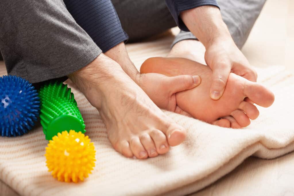
There is no question that foot pain can put a damper on the day to day. You might notice it in the morning as you get out of bed, after you have been on your feet for a long walk, or simply from wearing a particular pair of shoes. The cause is not uniform; an ache in the heel, arch, toes or the front and middle of the foot could be indicative of entirely different matters requiring their own treatment.
Given that the feet take a good deal of force with each step, minor things have a way of compounding. It is therefore worth paying attention to the nature of the discomfort and what aggravates it. Such observations will inform whether a change of footwear and some rest are in order, or if a consultation with an orthopaedic specialist is called for.
Foot Pain Often Follows a Pattern
What You Should Know
Pain in different parts of the foot can suggest different underlying problems.
Heel pain is often linked to plantar fasciitis, especially when pain is worse with your first steps in the morning.
Pain near the big toe may be caused by a bunion, arthritis, or a painful inflammatory condition such as gout.
Burning, tingling, or numbness can indicate nerve-related problems rather than joint or tendon issues.
Toe pain may come from deformities like hammertoes, ingrown toenails, arthritis, or injury.
Arch pain can be related to overuse, strain, tendon problems, or changes in foot shape.
Sudden swelling, redness, or severe tenderness can be a sign of inflammation, infection, or another urgent condition.
Supportive shoes, activity changes, and early evaluation can often help stop symptoms from getting worse.
If pain lasts, affects how you walk, or follows an injury, it should be checked.
#image_title
Why Foot Pain Can Feel So Different From One Person to Another
With its array of small bones, joints and soft tissues, not to mention ligaments, tendons and nerves, the foot is a complex structure. The proximity of all these elements means there are any number of ways for pain to set in. And since some of the culprits can present in much the same way at first, one has to be particular about the details.
Where the pain is situated is telling; what is felt in the heel will have different origins than trouble in the toes or ball of the foot. It is useful to note if the discomfort is sharp or dull, does it radiate, and which activities serve to aggravate or relieve it.
A Quick Look at Where Foot Pain Happens
Location of Pain
Possible Causes
Common Clues
Heel
Plantar fasciitis, heel pad irritation, stress injury
Pain with first steps, soreness after standing or walking
Arch
Strain, tendon irritation, overuse
Aching through the middle of the foot, worse with activity
Ball of foot
Metatarsalgia, nerve irritation, overload
Burning or pressure under the front of the foot
Big toe joint
Bunion, arthritis, gout
Bump, stiffness, swelling, redness, pain with push-off
Toes
Hammertoe, ingrown nail, arthritis, fracture
Crooked toe shape, shoe pressure, swelling, tenderness
Plantar fasciitis is a frequent culprit when it comes to heel pain. The condition has its origins in the plantar fascia, that thick band of tissue running along the sole of the foot which is responsible for arch support. Irritation of this tissue is enough to make standing or walking uncomfortable.
Those suffering from it will typically feel a sharp, stabbing sensation at the base of the heel. It is most pronounced with the first few steps out of bed in the morning or one gets up after being seated for some time. While the discomfort might subside with a bit of movement, it has a way of coming back once there has been prolonged standing or walking.
Pain is usually felt at the underside of the heel
Symptoms are often worst when you first stand up
Long periods on your feet can make it worse
Unsupportive shoes may increase discomfort
Pain in the Ball of the Foot
An overloading of the metatarsal bones and the soft tissues in their vicinity can give rise to pain at the front of the foot. It is an area that endures considerable stress from activities like running or walking, or even just standing for long periods, particularly when one is not wearing shoes with adequate support.
The way it is experienced varies; some will have a bruised or aching feeling while for others there may be a burning, tingling or numbness indicative of nerve irritation. Take these symptoms seriously if they keep getting worse.
Discomfort may feel like you are walking on a pebble
Pain may worsen in narrow or high heeled shoes
Numbness or burning can suggest nerve involvement
Big Toe Pain, Bunions, and Joint Problems
It is not unusual to experience pain in the big toe joint, and there are a number of possible origins for it. Take the bunion, for instance: as the joint at the base of the toe goes out of alignment, a bump forms that is plain to see and can get irritated from shoe friction.
Then there is arthritis, which will make the joint stiff and swollen, causing discomfort when one puts weight on it while walking. The area can turn red and hot, becoming extremely tender. Gout is one form of inflammatory arthritis that has a way of coming on with little warning.
Bunions often cause a bump and pressure from footwear
Arthritis may limit how well the toe bends
Gout often causes sudden pain, swelling, and redness
Toe Pain and Toe Deformities
There are a number of possible causes for toe pain, from injury and nail issues to arthritis or hammertoes. The latter is an abnormal bending of the toe at a joint that will put pressure on the foot and cause it to rub and hurt inside one’s shoes.
Then there is the matter of ingrown toenails; these can be quite painful and often result in some redness and swelling at the nail’s edge. And should a toe swell up after being stubbed or following a fall, it would be prudent to have any sprain or fracture ruled out.
Arch Pain and Mid foot Discomfort
With the arch serving to take the shock and spread out the weight, any strain there is bound to make walking a discomfort. One might feel pain in the middle of the foot or through the arch as a result of overuse, an irritated tendon or some alteration in the way the foot bears the body’s load.
Such pain does not always come on all at once; it can be a gradual thing. Then again, it may set in after an active day on one’s feet, a shift in routine or from shoes lacking in proper support. It is well to note these kinds of triggers when it comes to treatment.
When Nerves May Be Involved
The source of foot pain is not always the bones or joints. When one experiences a burning, tingling or numbness, or something akin to an electric shock, it can be indicative of an underlying nerve issue. Such sensations will make themselves known in various parts of the foot, from the toes and ball of the foot to more extensive areas.
It is worth giving these symptoms some consideration, particularly should they begin to worsen, alter your gait or put your balance at risk.
Signs You Shouldn’t Ignore
For the most part, self-care is a suitable course of action when one is dealing with mild symptoms that are likely to pass in short order. Should any red flags make an appearance, however, it is wiser to seek a medical evaluation without delay. A prompt diagnosis will do much to address problems like infection, fracture, gout, or a blood flow issue.
Pain that does not improve with rest, or keeps coming back
Swelling that keeps increasing or becomes severe, especially if it happens quickly
Spreading redness or skin that feels warm and tender, especially if you also have a fever
Inability to bear weight
Pain that starts suddenly and becomes very severe
Numbness or loss of sensation
A cold, pale, or blue foot or toes
Open wounds, blisters, puncture marks, or any drainage
Signs that infection may be present, such as warmth, increasing pain, redness, fever, or pus-like drainage
Recent injury, a twist, or a fall followed by worsening pain or swelling
Symptoms in a person with diabetes, neuropathy, immune suppression, or poor circulation
A change in foot shape or toe position after injury
Common Factors That Can Make Foot Pain Worse
Many foot conditions are made worse by repeated stress and poor support. Even when the cause is different, the same daily habits can keep symptoms going. Small changes can help, but they should not delay care when red flags are present.
Wearing shoes that are tight, worn out, or unsupportive
Suddenly increasing walking, running, or other exercise
Standing for long hours on hard surfaces
Ignoring pain and continuing activities that clearly make it worse
Changes in foot alignment or toe shape over time
What You Can Do at Home
For a case of mild foot pain, home remedies are an option as one monitors for any betterment. The approach taken ought to be in keeping with the nature of the symptoms and how the discomfort came about. But there is no place for home care when red flags are present; do not use home care if you have red flags such as inability to bear weight, rapidly increasing swelling, or signs of infection.
Rest from activities that clearly make the pain worse
Choose shoes with better cushioning and support
Avoid tight shoes if you have bunion irritation or toe pain
Use ice if the area feels sore or swollen
Track whether symptoms are improving, staying the same, or spreading
When to See an Orthopaedic Specialist
It is advisable to have foot pain seen to if there is no sign of improvement, or should it alter your gait or follow an injury. Left to its own devices, such pain can be persistent and lead to compensatory habits that put the back, hip, knee or ankle under strain.
An orthopaedic assessment will determine if the problem lies in the soft tissues, nerves, tendons, joints or bone. With the root cause established, one can then put in place a treatment plan suited to the condition rather than merely addressing the symptom.
When Foot Pain Needs Medical Attention
There are certain causes that call for a timely diagnosis and course of treatment, not least gout, infection or fractures. Vascular issues and more serious forms of inflammation fall into this category as well. In the event of any red flags, or should symptoms be on the upturn, it is advisable to seek medical counsel without delay.
Situation
Why It Matters
Next Step
Pain lasts more than a few days or keeps returning
May suggest an ongoing structural or inflammatory problem
Schedule an evaluation
You cannot bear weight
Could indicate a more significant injury or other urgent issue
Seek prompt care
Severe or rapidly worsening swelling
May signal inflammation, injury, or infection that needs fast help
Get medical guidance
Spreading redness or warmth, especially with fever
May suggest infection or inflammatory flare
Get medical guidance soon
Open wounds or drainage
May indicate infection
Seek prompt care
Numbness, tingling, or loss of sensation
May suggest nerve involvement or another urgent problem
Have it assessed
Pain began after a twist, fall, or direct injury
A sprain or fracture may need treatment
Seek orthopaedic evaluation
Symptoms in diabetes, neuropathy, immune suppression, or poor circulation
Higher risk of complications
Seek prompt medical advice
Cold, pale, or blue foot
May signal circulation problems
Get urgent evaluation
Getting the Right Diagnosis Matters
One can be forgiven for thinking foot ailments are all the same, yet the right course of treatment is a matter of getting to the root of it. Heel pain does not automatically mean plantar fasciitis, nor is every case of toe pain a bunion. Even a burning sensation may have nothing to do with the fit of one
u2019s shoes. An accurate diagnosis is what will point the way to the safest approach.
Should the pain in your feet get in the way of your day, an evaluation is in order to determine the source and the best remedy. There is much to be said for early intervention; in many cases, early care can reduce pain and lower the chance of a small issue turning into a longer-term problem.
Schedule an exam if your foot pain is persistent, worsening, or making it hard to stay active comfortably, or if you have any red flags like inability to bear weight or suspected infection.
This blog post is meant to be informative and should not act as a self-diagnosis tool. If you’d like to see one of our doctors, please contact us here.
Numbness and Tingling in Your Hands: What It May Mean
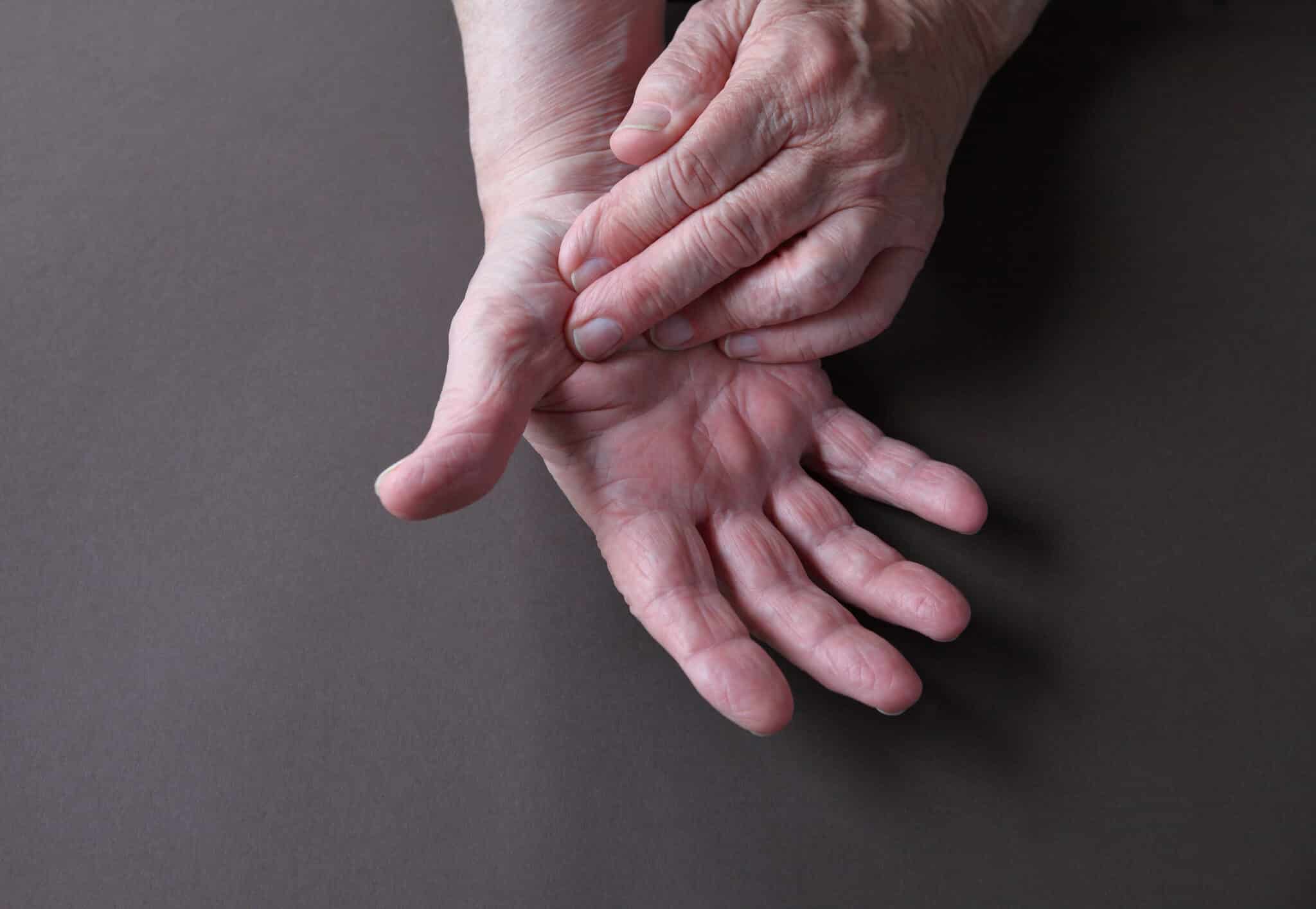
It is all too easy to put down a bit of numbness or tingling in the hands, particularly when it is intermittent. But if the sensation persists to the point of waking you at night, or you find your grip is not what it should be, there is a good chance a nerve is being compressed or otherwise irritated.
One might feel pins and needles in the course of everyday activities like typing, driving or even holding a phone. For some, there is an accompanying burning pain or weakness that can radiate up the forearm or into the elbow and wrist. Paying attention to such things will give you some idea of where the trouble lies.
Since similar symptoms have different origins, it is better to have an exam than to make assumptions. A clinician can determine where the nerve signals are off and put you on a course of treatment suited to the underlying cause. Being aware of the usual culprits is a good way to know when professional care is warranted.
#image_title
Common Reasons Your Hands May Feel Numb or Tingly
What You Should Know
Numbness and tingling often happen when a nerve is under pressure somewhere between your neck and your hand.
Carpal tunnel syndrome is a common cause, but it is not the only one.
Cubital tunnel syndrome can affect the ring and small fingers, especially when your elbow stays bent for long periods.
Symptoms may show up during sleep, computer work, driving, or repeated hand use.
Weakness, dropping objects, or trouble with fine tasks can suggest the nerve is affected more than before.
Some people feel symptoms in all fingers, while others notice it in certain parts of the hand.
Neck problems can also cause numbness or tingling that travels down the arm into the hand.
Getting help early may reduce irritation before lasting nerve changes develop.
Splints, activity changes, therapy, medicines, or surgery may be considered based on the cause.
When symptoms keep returning, it is safer to get them checked rather than waiting for them to worsen.
Why These Symptoms Happen
The nerves are responsible for relaying signals from the brain and spinal cord to the rest of the body. Should a nerve be compressed, inflamed or otherwise irritated, those signals will be altered. The result can be weakness, a burning pain, or a sense of numbness and tingling.
Symptoms themselves are often telling. One might look at which fingers are affected, what time of day the issue presents itself, or if there is accompanying pain in the elbow, wrist or neck to determine where the nerve is being impacted.
Frequently the problem is one of compression; as the nerve passes through some of the body’s more confined spaces, pressure builds on it. Yet that is not the only explanation, making a proper evaluation necessary.
Carpal Tunnel Syndrome
When one experiences numbness or a tingling sensation in the hand, carpal tunnel syndrome is a frequent culprit. The cause is compression of the median nerve in its passage through the carpal tunnel, that constricted area of the wrist.
This condition often affects the thumb, index finger, middle finger, and part of the ring finger. In the early stages the problem may be intermittent, but as time goes on the symptoms will tend to make themselves felt with greater regularity.
Numbness or tingling in the thumb, index, and middle fingers
Symptoms that are worse at night or when you first wake up
Discomfort during driving, typing, or holding a phone or book
Hand weakness or trouble gripping objects
Occasional dropping of items unexpectedly
Because carpal tunnel syndrome may slowly worsen, it is a good idea to seek care if symptoms are becoming more frequent or if weakness begins. Early steps may help reduce irritation.
Cubital Tunnel Syndrome
The ulnar nerve is the one that gives you a jolt of electricity when you put your elbow on something and hit your “funny bone.” It is this same nerve, running down the inside of the elbow, that is involved in cubital tunnel syndrome.
Irritation or compression of the ulnar can result in a tingling or numbness in the small and ring fingers. One will typically find the symptoms are more pronounced after the elbow has been held in a bent position for any length of time, be it from working at a desk or while sleeping.
Tingling in the ring and small fingers
Symptoms that show up when sleeping with the elbow bent
Numbness during phone use or desk work
Hand weakness, especially with pinch or grip tasks
Clumsiness with fine motor tasks
Could the Problem Be Coming From Your Neck?
The source of nerve trouble in the hand is not always to be found in the hand or wrist itself. At times, an irritated nerve root in the cervical spine, that is to say the neck, will send its way down the arm and into the hand.
Such a condition is known as cervical radiculopathy. One might experience neck pain or some discomfort in the shoulder, in addition to the usual numbness or tingling, with pain even radiating down the arm. All of these are telling signs for a proper evaluation.
Common Patterns of Hand Numbness and Tingling
Possible Cause
Where You May Feel It
Common Clues
Carpal tunnel syndrome
Thumb, index, middle, and part of the ring finger
Often worse at night, during driving, typing, or gripping
Cubital tunnel syndrome
Ring finger and small finger
Often worse with a bent elbow, sleep, or phone use
Neck-related nerve irritation
Can travel down the arm into the hand
May happen with neck pain, shoulder pain, or radiating arm symptoms
Other Symptoms to Watch For
Numbness and tingling can be an early warning sign, but they are not always the only symptoms. As nerve irritation continues, other problems may appear and can help show how much the nerve is being affected.
Burning or aching pain
Weakness in the hand or fingers
Difficulty gripping, pinching, or buttoning clothing
Dropping objects more often
Symptoms that wake you from sleep
If you are noticing weakness or losing hand control, it is especially important not to wait too long for an evaluation. That can help reduce the chance of lasting problems.
How We Evaluate Hand Numbness
We will begin the exam by going over your symptoms and an inspection of the neck, shoulders, elbows, wrists and hands. The way the numbness has been presenting itself, its duration and what seems to set it off are all useful in determining where to go from there.
The clinician is likely to test reflexes as well as skin sensation and strength. Should it be necessary to see just how badly a particular nerve is being affected and make a positive identification, further testing might be put forward.
#image_title
Treatment Options
Treatment depends on the cause of your symptoms, how long they have been present, and whether nerve weakness is involved. Many people improve with non-surgical care, especially when treatment starts sooner.
Activity changes to reduce pressure on the nerve
Bracing or splinting, especially at night for certain conditions
Anti-inflammatory medication for pain or swelling when it is appropriate for you
Physical therapy or occupational therapy to improve movement and function
Surgery for ongoing compression or progressive weakness in selected cases
While anti-inflammatory drugs can be of service in reducing pain and inflammation, they are not a cure for the underlying nerve compression. Nor should they be seen as a substitute for an accurate diagnosis. For some, carpal tunnel symptoms will abate with such medication, but it is unlikely to be sufficient on its own.
Safety: When to Get Emergency Help
Should you experience a sudden onset of weakness or numbness in an arm or hand, particularly on one side, emergency medical attention is warranted. Do not delay in getting help if such symptoms are accompanied by any of the following: facial drooping, difficulty with speech, dizziness, confusion, a loss of balance, chest pain, shortness of breath or a bad headache.
Fast treatment is necessary as these may be signs of a stroke or some other pressing condition. When in doubt, the prudent course of action is to contact emergency services or make for the closest emergency department.
Note: Not All Hand Numbness Comes From Nerve Compression
Hand numbness can have medical causes beyond pressure on a nerve. Possible causes include diabetes-related neuropathy, vitamin B12 deficiency, thyroid disease, medication side effects, alcohol-related neuropathy, inflammatory or autoimmune neuropathies, and circulation-related problems. A checkup can help sort out the right reason.
When It’s Time to See a Specialist
Consider scheduling an evaluation if symptoms keep coming back, interrupt sleep, or start affecting daily activities. Ongoing numbness and tingling should not be ignored, especially if they come with weakness or trouble using your hand.
Your symptoms keep returning
You wake up with numb or painful hands
You have weakness or trouble gripping objects
Your symptoms move up the arm or start in the neck
Daily tasks like typing, driving, dressing, or cooking are becoming harder
The right diagnosis can help you avoid making nerve irritation worse and can point you toward treatment that matches the true cause.
Don’t Ignore Persistent Hand Numbness
When one experiences recurring pain, weakness, or a sensation of numbness and tingling, it is often an indication of a nerve problem that warrants some attention. An early assessment is the best way to safeguard function and make a return to daily life more comfortable, no matter if the trouble appears to be in the neck, elbow or wrist.
Should the symptoms in your hands fail to abate or begin to get in the way of things, we would suggest an appointment with Princeton Orthopaedic Associates for a proper examination.
This blog post is meant to be informative and should not act as a self-diagnosis tool. If you’d like to see one of our doctors, please contact us here.
Ice or Heat for Pain Relief: How to Know Which One to Use
There is a tendency for people to put an ice pack or heating pad on when they are in pain or sore, not always sure which is the better remedy. While both have their uses, they go about it in different ways. As a rule of thumb, ice is preferable for fresh injuries where there is swelling, whereas heat will do more for tightness in the muscles or general stiffness.
Applied properly, either can be an aid to comfort and mobility as well as for managing swelling and pain. One should also be mindful of using them in a safe manner. Should the symptoms prove to be severe, fail to get any better or come back time and again, it would be prudent to seek medical counsel.
Quick Differences Between Ice and Heat
Use Ice When You Want to Calm Things Down
Often best after a recent injury
May help reduce swelling
Can ease pain by temporarily numbing the area
Common choices for sprains, strains, and bruises
Use Heat When You Want to Loosen Things Up
Often helpful for stiffness and tight muscles
May increase blood flow to the area
May improve flexibility before movement
Common choices for chronic aches and muscle tension
How Ice Helps an Injured Area
Applying cold therapy is a way to put the brakes on blood flow in the affected region, which can be of some assistance in settling any swelling or irritation in the wake of an abrupt injury. The ice will also serve to dull the pain by numbing sore tissue for a time. While it is not going to remedy the underlying problem on its own, it does allow for more comfortable movement and rest.
One will typically turn to ice in the immediate aftermath of an impact, sprain or strain. This is particularly true if there is throbbing pain, warmth, bruising or visible swelling. A hard bump that has begun to mark the skin, a fresh knee sprain or even a rolled ankle would be typical cases for its use.
How Heat Helps Sore or Stiff Tissue
Applying heat can ease tight muscles and may improve circulation to the part that is sore. The result, for many people, is a stiff joint that can feel more at ease and yield to movement with less difficulty. When the pain is of an achy, tight nature as opposed to being sharp and swollen, many will find warmth to be of particular use.
It is a good remedy for persistent muscle soreness or stiffness in the back and neck where there is no obvious swelling. One might also turn to heat prior to some light activity or a bit of gentle stretching; it can help movement feel less stiff.
A Simple Way to Decide
For a fresh problem accompanied by swelling, warmth or an outbreak of pain, ice is the way to go. On the other hand, where stiffness and tightness are the chief complaints and there is little to no swelling, heat will generally be more effective for the ache. One should observe the area and make any adjustments with care.
While this is not a rule that applies to every injury or constitution, it is sound advice for most ordinary orthopedic discomforts. In cases of doubt, stick to safe measures and if there is no sign of improvement, have the matter evaluated by a professional.
When Ice Is Usually the Better Choice
Right after a sprain or strain
When a joint or muscle is swollen
For bruises or impact injuries
After activity if the area looks more inflamed
When pain feels sharp, hot, or throbbing
When Heat Is Usually the Better Choice
For muscle tightness or spasms
For stiffness in the neck, back, shoulder, or other joints
Before stretching or gentle exercise
For ongoing aches that are not swollen
When movement feels limited because tissue feels tight
Ice vs Heat at a Glance
Situation
Usually Try
Why It May Help
Recent sprain or strain
Ice
May help reduce swelling and numbing pain
Visible swelling
Ice
Cold can calm inflammation in the area
Bruise or bump
Ice
Often used early for pain and swelling
Muscle tightness
Heat
Warmth can help muscles relax
Joint stiffness
Heat
Heat can make movement feel easier
Soreness after activity with swelling
Ice
Often better when inflammation is present
Ongoing achiness without swelling
Heat
Warmth may improve comfort
Can You Alternate Ice and Heat?
There is a time and place for both cold and warmth, particularly as one’s symptoms go from swelling to stiffness. Take an injury for instance: ice may be the way to go in the beginning, but once the swelling has subsided and you are left with tightness, heat will likely prove more soothing. Just be sure to observe proper timing and discontinue if there is any aggravation of symptoms.
Should you be uncertain of where you stand, let the signs tell you. A good rule of thumb is that ice is indicated when there is swelling and heat, whereas a lack of swelling accompanied by stiffness suggests heat would be better. If any doubt remains, err on the side of caution and seek some advice.
How to Use Ice Safely
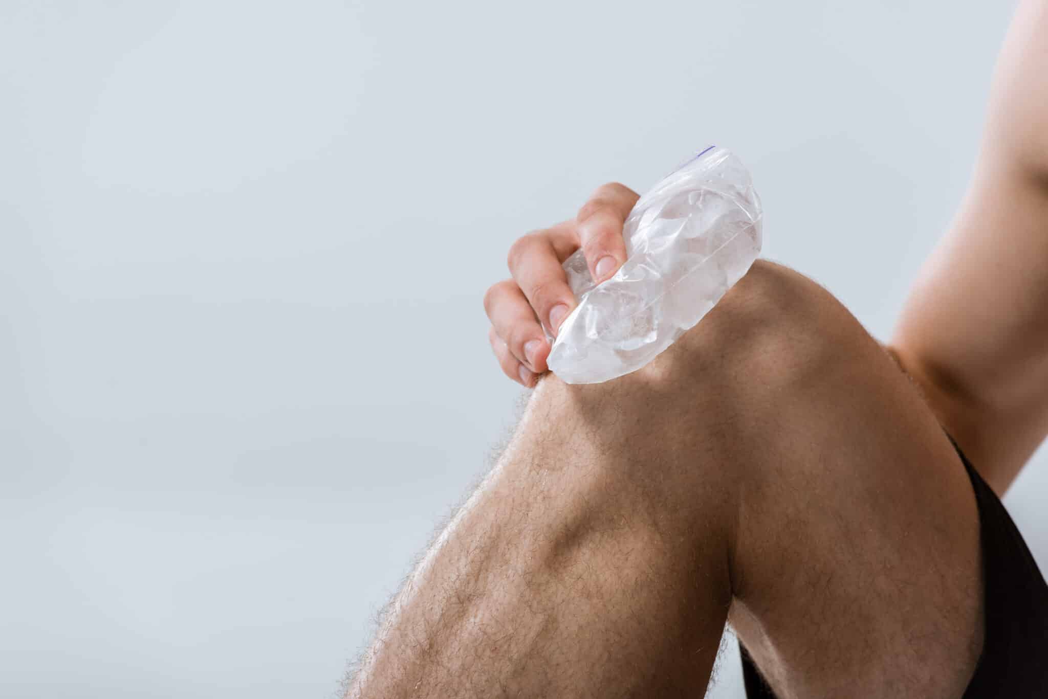
Wrap an ice pack or bag of ice in a towel before applying
Do not place ice directly on skin
Use for about 15 to 20 minutes at a time, then take a break
Check the skin during and after treatment
Stop if skin becomes painful, blotchy, white, blistered, unusually red, feels burned, or if the area stays numb after treatment
Do not use ice over broken skin or areas with poor circulation unless a clinician tells you to
How to Use Heat Safely
Use a warm heating pad, warm towel, or another gentle heat source
Heat should feel warm, not hot or scalding
Use for about 15 to 20 minutes at a time while checking the skin
Stop if skin becomes painful, blotchy, white, blistered, unusually red, feels burned, or if the area remains numb after treatment
Avoid heat on new injuries, active swelling, redness, warmth, infection, or bleeding
Avoid falling asleep with a heating pad on
When Heat or Ice Should Be Used With Extra Caution
Those with reduced sensation, diabetes, neuropathy or other nerve issues stemming from the condition should consult a clinician prior to applying ice or heat. The same goes for anyone with poor circulation, peripheral vascular disease, Raynaud’s, or simply an inability to gauge temperature properly. One can easily do damage to the skin with cold or heat and not be the wiser for it, so some additional precautions are in order.
Avoid heat on any part of the body that is red, hot, swollen or otherwise inflamed, especially after a new injury. Heat is out of the question if there is a fever, infection or bleeding. As for open wounds or any skin that is not in good shape, ice should not be put on it without the go ahead from a clinician.
When to Get Checked by an Orthopedic Specialist
While ice and heat are useful for easing pain, putting swelling in check and allowing for more comfortable movement, they are no substitute for a proper evaluation if something more serious is at play. Should the pain be considerable, fail to improve, or change for the worse, particularly following an injury, it is best to have it examined.
There are certain warning signs that call for a trip to urgent care or other timely medical intervention: deformity, a suspected fracture, or an inability to put weight on the limb. The same goes for severe or escalating pain, new weakness of any kind, numbness, tingling, a fever or any indication of infection.
Possible blood clot symptoms also need urgent attention. Watch for calf swelling, warmth, redness and pain, especially if it happens in one leg or does not improve. Do not make the mistake of using ice or heat in place of getting the care you need when such symptoms present themselves.
Schedule an Exam if Pain Isn’t Improving
There are times when it is not clear if ice or heat is the right course of action for one’s symptoms, or when pain simply will not abate. In such cases, Princeton Orthopaedic Associates is on hand to assist. We can put you through a clinical evaluation to get to the bottom of the discomfort and recommend a more appropriate treatment plan that is in keeping with your particular injury and what you want to achieve in your recovery.
This blog post is meant to be informative and should not act as a self-diagnosis tool. If you’d like to see one of our doctors, please contact us here.
Hip Pain While Sitting: Common Causes and Ways to Find Relief
There is a certain frustration to having hip pain when one is seated; it can put a damper on work, meals, driving and any time off. The root of the problem is frequently an irritation of the muscles, tendons, joints or nerves in the vicinity, and the way the symptoms present themselves will often tell you something about what is at play.
It could be an ache along the side of the hip, a pinching sensation in the groin, stiffness upon rising, or discomfort that radiates down into the thigh or buttock. While some find relief through a bit of home care and by adjusting their posture and activities, any pain that lingers or gets worse should be properly assessed in order to get the appropriate treatment underway.
Key Points About Hip Pain During Sitting
What You Should Know
Pain while sitting may come from the hip joint itself or from muscles, tendons, bursae, or nerves around the hip.
The location of the pain matters. Groin pain often points toward the hip joint, while pain on the side or back of the hip may suggest another source.
Long periods of sitting can increase pressure on irritated tissues and tighten the muscles that help support the hip.
Simple steps, like changing position, standing up often, and using supportive seating, may help reduce symptoms.
Common causes include bursitis, arthritis, muscle or tendon irritation, a hip labral tear, nerve-related pain, and referred pain from the lower back.
Ice, gentle stretching, and changes in activity may help in early or mild cases.
Persistent pain, pain that affects walking, or pain accompanied by weakness, numbness, or catching should be evaluated by a specialist.
Treatment depends on the cause and may include physical therapy, medications, guided exercise, or other orthopaedic care.
Why Sitting Can Make Hip Pain Worse
Prolonged sitting keeps the hips in a bent position, which is apt to put added pressure on the hip joint and its environs. For soft tissues that are stiff or already in an inflamed state, this can be a source of strain.
Then there is the matter of inactivity. A lack of movement while you are seated will cause the muscles about the hip to become rigid over time. The result is that any discomfort makes itself known all the more when one gets up to walk or simply changes position.
Where You Feel the Pain Can Help Identify the Cause
The exact location of your pain can help narrow down possible causes. Hip pain is not always felt directly over the joint, and different structures tend to cause pain in more specific areas.
Groin or front of the hip: often linked with the hip joint, such as arthritis or a labral tear.
Outer side of the hip: often related to bursitis and/or irritation of the gluteal tendons, which is frequently grouped under greater trochanteric pain syndrome.
Buttock or back of the hip: may involve deep hip muscles, the sacroiliac area, or pain that travels from the low back.
Pain into the thigh or below the knee: can happen with nerve irritation or referred pain from the spine.
Common Causes of Hip Pain While Sitting
Many conditions can cause hip discomfort in a seated position. Some affect the joint itself, while others involve soft tissue around the hip. Some problems start in the lower back and are felt near the hip.
Hip osteoarthritis
Osteoarthritis is the wear and tear of the joint cartilage. It often causes stiffness and aching in the groin or thigh. Pain may feel worse after sitting for a while or when you first get up.
Hip bursitis
There are small sacs called bursae, filled with fluid to keep friction at bay between tissues in motion. Should a bursa get irritated, it can be the source of pain on the outside of the hip, particularly when one is sitting or has to lie on that side. Then again, such discomfort in the outer hip might stem from the gluteal tendons; this type of irritation is commonly referred to as greater trochanteric pain syndrome.
Muscle or tendon irritation
Tight or overused muscles and tendons around the hip can become sore, particularly after exercise, repetitive activity, or prolonged poor posture. This can lead to a deep ache, tightness, or a pulling feeling while seated.
Hip labral tear
The labrum is a ring of cartilage that helps stabilize the hip socket. A tear can cause groin pain, clicking, catching, or a sense that the hip is not moving smoothly. Sitting can increase discomfort for some people.
Sciatica or other nerve-related pain
If a nerve is irritated, pain may start in the buttock or low back and travel into the hip or leg. Sitting can worsen these symptoms, especially when numbness, tingling, or burning is also present.
Referred pain from the lower back
Hip pain does not always come from inside the hip. Problems in the lower spine can create pain that feels like it is centered in the hip, especially when sitting for long periods.
How Different Causes of Sitting-Related Hip Pain May Feel
Possible Cause
Common Pain Location
What You May Notice
Hip osteoarthritis
Groin, front of hip, thigh
Stiffness, pain after sitting, pain when first standing up
Hip bursitis
Outer side of hip
Tenderness, pain with pressure, discomfort when sitting or lying on one side
Muscle or tendon irritation
Front, side, or buttock area
Ache, tightness, soreness after activity or prolonged sitting
Labral tear
Groin or deep hip
Clicking, catching, pinching, painful sitting
Nerve-related pain
Buttock, hip, leg
Burning, tingling, numbness, pain that travels
Referred pain from low back
Back of hip or buttock
Sitting worsens symptoms, back pain may also be present
What You Can Do at Home to Ease Hip Pain
For mild symptoms with no history of a recent fall or other trauma, there are some straightforward measures one can take at home to ease the irritation. What is important is to lessen the strain without allowing the hip to become stiff. Should any of these approaches aggravate the pain, they ought to be discontinued.
Change positions often instead of sitting for long stretches.
Stand up and walk briefly every 30 to 60 minutes if you can.
Use ice for short periods if the area feels inflamed or sore after activity.
Try gentle stretching only if it does not increase pain. Stop if it worsens your symptoms.
Check your chair height and support so your hips and knees feel comfortable.
Avoid sitting on very hard surfaces if that clearly worsens symptoms.
When taking an over the counter medication, be sure to follow to what the label says as well as any instructions you may have gotten from a doctor. In the case of anti-inflammatory drugs, it is best to seek some direction from a professional first should you have kidney trouble, be on blood thinners or are pregnant. The same applies if there is a history of stomach ulcers or bleeding, or any other medical issues to consider.
Helpful Sitting and Movement Adjustments
Small changes in how you sit and move during the day can help. These strategies may be most helpful when pain is related to posture, soft tissue irritation, or joint stiffness. If you notice worsening pain, scale back and consider getting evaluated.
Sit with your feet flat on the floor and avoid crossing your legs for long periods.
Keep your lower back supported so you are not slumping forward.
Use a cushion or supportive seat if hard chairs increase pain.
Break up long drives with standing or walking stops when possible.
Return to exercise gradually and avoid pushing through pain that is increasing.
When Hip Pain While Sitting Should Be Evaluated
Some hip pain improves with time and careful self-care. Yet one should not assume that pain in the hip from sitting is nothing at all. There are occasions when it is more than something simple. Any symptoms that do not go away, or those that are on the increase, as well as pain following an injury or indications of a nerve or systemic issue, should warrant an evaluation to get to the root of the problem and put in place the right treatment.
Pain lasts more than a few days or keeps coming back.
The pain is getting worse instead of improving.
You are limping or changing how you walk.
You notice catching, locking, or clicking in the hip.
You have numbness, tingling, or pain that travels down the leg.
The pain interferes with work, sleep, exercise, or daily activities.
When to seek more urgent care
You can’t bear weight on the leg.
The pain began after a fall or other injury.
Fever or chills are present.
You notice redness or warmth around the hip.
Inability to move the hip is present.
Visible deformity is present.
Severe pain at rest or at night is present.
Unexplained weight loss is present.
There is a history of cancer or a high risk of infection.
Loss of bowel or bladder control is present.
Saddle numbness is present.
Rapidly worsening weakness, numbness, or tingling is present.
You have significant weakness, sudden swelling, or severe pain.
How Orthopaedic Specialists Diagnose Hip Pain
An examination is almost always the first step in making a diagnosis. From there, we can determine the source of the pain and what kind of movements set it off, as well as assess hip function and any possible back involvement.
If the symptoms require it, you may be advised to have imaging done, be it an X-ray or another form of study. Such tests are useful for spotting structural issues, arthritis or other ailments that are beyond what home remedies can address.
Treatment Options for Ongoing Hip Pain
Treatment depends on the source of the pain. Many patients improve with non-surgical care aimed at reducing irritation, improving movement, and strengthening the muscles that support the hip.
Activity modification to reduce pain triggers
Physical therapy to improve flexibility, strength, and movement mechanics
Anti-inflammatory medication when appropriate
Image-guided injections for certain diagnoses
Further orthopaedic evaluation for structural hip problems or advanced arthritis
If symptoms are not improving, getting the right diagnosis early may help reduce prolonged pain and unnecessary limit setting.
Don’t Ignore Hip Pain That Keeps Returning
You should not have to live with hip pain when you sit. The nature of the symptoms will point to what is required for some relief, be the source the joint itself, soft tissue in the vicinity or the low back. If the pain lingers, gets worse or affects your day to day life, please call your doctor or schedule an appointment with the POA Team.
This blog post is meant to be informative and should not act as a self-diagnosis tool. If you’d like to see one of our doctors, please contact us here.
When you break a bone, twist an ankle, or wake up with a joint too swollen to move, you have two obvious options: sit in a hospital emergency room for hours, or wait days for a specialist appointment. There's a better third option most New Jersey families don't know exists. Princeton Orthopaedic Associates offers walk-in orthopedic urgent care at multiple locations across central New Jersey, staffed by the same orthopaedic specialists our patients have trusted for over 40 years. No appointment needed, no ER wait times, and you're seen by providers who treat bones, joints, and muscles all day, every day. In this post, we'll cover what orthopedic urgent care is, which injuries it's designed for, why it often beats the emergency room for musculoskeletal injuries, and exactly what to expect when you walk into a POA urgent care location.
What Is Orthopedic Urgent Care?
Orthopedic urgent care is walk-in medical care focused specifically on injuries to your bones, joints, muscles, tendons, and ligaments. Think of it as the middle ground between a hospital emergency room and a scheduled specialist visit. At POA's orthopaedic urgent care, you're evaluated by orthopaedic providers with on-site X-ray imaging, so a suspected fracture can be diagnosed, casted or splinted, and given a follow-up plan in a single visit. If you or a member of your family has had an orthopaedic injury within the last 36 hours, you can walk in without an appointment and be seen by our team. It's the same practice, the same specialists, and the same standard of care our patients have relied on across central New Jersey, just available when injuries actually happen: evenings, weekends, and everything in between.
Orthopedic Urgent Care vs. the Emergency Room: Which Should You Choose?
For a true medical emergency, always call 911 or go to the nearest emergency room. Head injuries, uncontrolled bleeding, bones visibly protruding through the skin, or an injury combined with chest pain, difficulty breathing, or loss of consciousness all belong in the ER. But for the vast majority of orthopedic injuries, the emergency room means long waits, higher bills, and an evaluation by a generalist who will often simply refer you to an orthopaedic specialist anyway. Orthopedic urgent care flips that experience. At POA, you walk in and go straight to the specialist. Our providers diagnose your injury with on-site imaging, begin treatment immediately, and connect you directly to our full team of orthopaedic surgeons and specialists if your injury needs surgical evaluation or ongoing care. You avoid the hospital emergency department wait, and your entire care journey stays within one practice from the first X-ray to the final follow-up.
Where Can I Find Walk-In Orthopedic Urgent Care in New Jersey?
Princeton Orthopaedic Associates offers orthopaedic urgent care at convenient locations across central New Jersey, with weekday, evening, and Saturday hours. Our Plainsboro office offers urgent care Monday through Friday with extended evening hours midweek, plus Saturday morning hours. Our Hillsborough office offers urgent care Monday through Thursday with evening hours, Friday during the day, and Saturday mornings. Our Princeton office on Princeton Avenue offers Saturday morning urgent care as well. Because hours and locations can occasionally change, we recommend checking our urgent care page for current hours or calling us at (609) 924-8131 before heading in. You can also view all of our offices on our locations page. Wherever you are in the Princeton, Plainsboro, Hillsborough, Robbinsville, Monroe, or Ewing areas, expert orthopaedic care is close by when an injury strikes.
What Should I Bring to My Urgent Care Visit?
A little preparation makes your walk-in visit even faster. When you come to POA orthopaedic urgent care, bring your health insurance card, a driver's license or photo ID, contact information for your health insurance policy holder, a list of medications you're currently taking, and a list of any allergies. We accept most major insurance plans, including Aetna, AmeriHealth, Cigna, Horizon PPO and HMO, Medicare, Oxford, motor vehicle insurance carriers, and workers compensation. If you were hurt on the job, our team is experienced in treating work-related injuries and handling the associated workers compensation documentation. Not sure whether your plan is accepted? You can review our full list of accepted insurances or give us a call before your visit.
Why Fast Treatment for Orthopedic Injuries Matters
With musculoskeletal injuries, timing genuinely affects outcomes. An untreated fracture can begin healing in the wrong position. A dislocated joint left unreduced becomes harder to treat and more painful by the hour. Even a "simple" sprain, if ignored, can lead to chronic instability and repeat injuries down the road. Getting evaluated within the first 36 hours means your injury is diagnosed accurately from the start, immobilized or treated correctly, and placed on a recovery plan built by specialists. It also means faster pain relief, which matters when you're the one limping around the house. Early specialist treatment frequently shortens overall recovery time and reduces the risk of complications that require surgery later. In short: don't wait it out and hope for the best. Walk in and know for sure.
Orthopedic injuries never happen at convenient times, but getting expert care for them can be convenient. Princeton Orthopaedic Associates' walk-in orthopaedic urgent care gives central New Jersey families direct access to orthopaedic specialists, on-site X-ray imaging, and same-day treatment for sprains, strains, fractures, dislocations, and sports injuries, without an appointment and without the emergency room wait. If you or someone in your family has been injured within the last 36 hours, call us at (609) 924-8131, text us at (609) 757-9992, or simply walk in to one of our urgent care locations. And for ongoing orthopaedic needs, you can always schedule an appointment with our team. When it comes to your bones and joints, go straight to the specialists first.
Understanding Low Back Pain
There are few ailments as common as low back pain when it comes to the reasons for a visit to the doctor. For some, it is a mild strain that is over in short order; for others it becomes an ongoing issue that can disrupt one’s work, exercise routine and sleep. An understanding of what may be at the root of the problem and how to deal with it takes much of the guesswork out of the matter.
The lower back is by nature a complicated region where nerves, ligaments, muscles, joints, discs and bones all have to function in concert. With so many structures in close proximity, there are a number of ways for pain to set in. It may be confined to the back or make its way down into the leg, thigh or buttock.
While the majority of cases will resolve on their time with non-surgical treatment, there are occasions when a prompt examination is warranted. This is particularly true if the pain is severe or recurrent, or if one notices any warning signs like numbness or weakness.
Key Facts About Low Back Pain
What You Should Know
Low back pain is very common and can affect people of all ages.
The pain may come from muscles, joints, discs, nerves, or a mix of structures.
Many cases improve with relative rest from painful activity, physical therapy, and guided exercise.
Pain that travels down the leg may point to nerve irritation, often called sciatica.
Good posture, core strength, and safe body mechanics may help reduce future flare-ups.
Low back pain can be acute, meaning short term, or chronic, meaning it lasts much longer.
Imaging such as X-rays or MRI is not needed for every patient, especially early on.
Loss of bowel or bladder control, numbness in the saddle or groin area, or new or worsening leg or foot weakness may signal a serious neurologic condition and needs emergency or same-day medical evaluation.
Most people do not need surgery, but some conditions do need specialist evaluation.
Early evaluation can help when symptoms are severe, keep coming back, or are not improving as expected.
What Causes Pain in the Lower Back?
A sudden motion, a case of heavy lifting or repetitive strain will sometimes be the cause of low back pain, though in some instances there is no apparent trigger. For many, the problem lies with muscles and ligaments that have been overstretched or irritated; for others it is the discs, facet joints or nerves in the vicinity.
Then there are the effects of age. With time one may find the spine has lost some of its flexibility, the joints have turned arthritic and the discs have given up their height and water content. While such changes do not inevitably lead to pain, they can be a factor in the symptoms of certain patients.
It is important to take a thorough history and conduct a physical exam since different conditions can present in much the same way. One can glean useful information from the pattern of the pain, its duration or if it is radiating down the leg, all of which will inform what comes next.
Common Sources of Low Back Pain
Low back pain is often grouped by the structure that seems most involved. In some cases, more than one issue may contribute at the same time, which can change how symptoms feel and how they respond to care.
Possible Source
What It Means
Common Pattern
Muscle or ligament strain
Overstretching or irritation of soft tissues
Pain after lifting, bending, twisting, or overuse
Disc problems
The cushion between spinal bones becomes irritated or bulges
Back pain, sometimes with leg pain or numbness
Facet joint arthritis
Wear and tear in the small joints of the spine
Stiffness and pain with standing, twisting, or extension
Sciatica or nerve compression
A spinal nerve becomes irritated or pinched
Pain shooting into the buttock, thigh, calf, or foot
Spinal stenosis
Narrowing around the nerves in the spine
Leg pain or heaviness with walking or standing
Spondylolisthesis
One spinal bone shifts relative to another
Back pain, leg symptoms, or pain with activity
How Low Back Pain May Feel
The sensation can vary based on the cause. Some people feel a dull ache or tightness in the center of the lower back, while others notice sharp pain with certain moves. How pain changes during the day can also give clues.
Stiffness when getting out of bed or after sitting
Muscle spasms or a feeling that the back has “locked up”
Pain with bending, lifting, twisting, or standing upright
Aching that stays in the low back or buttock
Pain, tingling, numbness, or burning that travels down the leg
Weakness in the leg or foot in more serious nerve-related cases
When pain travels below the knee, especially with numbness or weakness, nerve involvement becomes more likely. That does not always mean the problem is severe, but it usually deserves a closer look from a clinician.
Acute Pain Compared With Chronic Pain
Doctors often describe low back pain based on how long it lasts. Using time frames helps guide care and sets more realistic expectations about recovery. It can also help decide when follow-up is needed.
Type
Time Frame
Typical Approach
Acute
Less than 4 weeks
Activity modification, pain relief, and a gradual return to movement
Subacute
4 to 12 weeks
Focused rehabilitation and closer follow-up if symptoms continue
Chronic
More than 12 weeks
A broader plan that addresses strength, flexibility, mechanics, and underlying causes
One might see acute pain come on from a strain or flare-up, and in many cases it will subside in short order. Chronic pain is a more complicated matter; there can be deconditioning at play, or arthritis, disc issues and recurrent nerve irritation to contend with. When the pattern is long standing, a strategy is called for that does not stop at mere pain management.
When to Seek Medical Attention Right Away
Most low back pain is not dangerous, but certain symptoms can point to a more serious problem. These warning signs should not be ignored, because they may need emergency care or evaluation the same day.
Loss of bowel or bladder control
New or worsening weakness in the leg or foot
Numbness around the groin or saddle area
Fever, chills, or unexplained weight loss with back pain
Pain after a major fall, accident, or other trauma
History of cancer, infection, or other medical conditions that raise concern
A serious neurologic condition could be at play if there is an inability to control the bladder or bowels, numbness in the groin or saddle region, or new or progressive weakness in the leg or foot. Such symptoms are cause for emergency care or a medical evaluation on the day they present. There are other warning signs that likewise demand to be seen without delay.
How Low Back Pain Is Evaluated
One typically starts the evaluation by talking with the patient to establish the history of the pain: its onset and location, what aggravates it, and if there is any radiation down the leg. The clinician then goes on to examine posture, strength, reflexes, sensation and range of motion in order to correlate the symptoms with a likely cause.
While imaging has its place, it is not something every patient needs. There are circumstances where it is warranted, however, such as in the event of trauma or when red flags appear. It would be called for if one suspects an infection, fracture or cancer, if neurologic deficits are on the increase, or simply if the condition has failed to show any improvement with time.
Test
When It May Be Used
What It Can Show
X-ray
If fracture, alignment issues, or arthritis are concerns
Bone structure and spinal alignment
MRI
If nerve symptoms persist or serious causes are suspected
Discs, nerves, soft tissues, and spinal canal narrowing
CT scan
Sometimes used when more bone detail is needed
Detailed images of bone anatomy
Physical exam
Often needed to guide care
Strength, motion, nerve findings, and pain pattern
Non-Surgical Treatment Often Works Well
Most people with low back pain improve without surgery. Treatment depends on the cause of symptoms, how long they have lasted, and whether nerves are involved. The goal is to reduce pain while helping you move more safely and comfortably.
Short-term activity changes to reduce strain
Physical therapy focused on flexibility, posture, and core strength
Heat or ice, depending on what feels best and when symptoms began
Anti-inflammatory or other pain-relieving medication when appropriate
Guided exercise to restore movement and prevent recurrence
In some cases, spine injections for ongoing inflammation or nerve pain
#image_title
Helpful Early Steps at Home
For those with only a mild degree of pain and no cause for alarm, there are some straightforward approaches that can be of assistance. One is to rest, though in this context it is best understood as refraining from whatever is aggravating the condition rather than spending an inordinate amount of time in bed. In fact, so long as you do not exceed what is tolerable, some measure of movement will often aid in the process of getting better.
Stay away from bed rest for long periods
Take short walks if walking feels comfortable
Use good lifting mechanics and avoid sudden twisting
Start gentle stretching or exercises only if they do not worsen symptoms
Seek care if pain is intense, persistent, or spreading into the leg
When Surgery May Be Considered
For the majority of low back pain, surgery is not the initial course of action. It is an option only if non-surgical methods have failed to offer sufficient relief and the symptoms can be traced to a particular structural issue. A clinician will be able to put the risks and anticipated benefits in perspective following an examination.
Certain fractures, infections, or other serious spine conditions
If surgery becomes part of the discussion, the decision should be based on your diagnosis, nerve findings, level of disability, and response to other treatments. The plan should match your symptoms and overall health.
How to Lower Your Risk of Future Back Pain
While it is not possible to forestall every case of low back pain, one’s daily routine can be of assistance. The root of many a flare-up is to be found in deconditioning or poor body mechanics, as well as the kind of repeated strain or prolonged inactivity that comes with sitting for too long. Over time, even minor adjustments will show their worth.
Keep your core and hip muscles strong
Stay active with regular walking or other low-impact exercise
Lift with your legs and avoid twisting under load
Change positions often if you sit for long stretches
Maintain a healthy weight when possible
Follow through with physical therapy exercises after symptoms improve
When It’s Time to See a Specialist
If your pain is severe, keeps coming back, or is not improving with time and conservative care, an orthopaedic or spine evaluation is in order. A specialist will be able to determine if what you are experiencing is due to a disc issue, muscle strain, arthritis, spinal stenosis or some form of nerve compression.
There is also a case for seeing a specialist when the pain has begun to alter your gait, put restrictions on your day to day routine or otherwise diminish your quality of life. The earlier the root of the problem is pinpointed, the better positioned one is to put a more focused course of action in place.
Find the Right Care for Low Back Pain
There is a certain stress to low back pain, particularly when it interferes with the ordinary business of the day. For most, things can be put right with an accurate diagnosis and a non-surgical approach that gives due consideration to one’s movement, strength and the health of the spine. Should the pain not abate on its own, an evaluation is in order to get to the root of the matter and see which course of action is most suitable.
This blog post is meant to be informative and should not act as a self-diagnosis tool. If you’d like to see one of our doctors, please contact us here.
Tailbone Pain Can Make Sitting, Driving, and Daily Life Uncomfortable
There is a certain difficulty to everyday tasks when one has tailbone pain, or coccydynia. Sitting at work, rising from the bed, even just leaning back in a chair can become trying. While it often follows an injury such as a fall or is a consequence of childbirth, there are times it will develop with no obvious cause.
The tailbone is the lowest part of the spine and what bears your weight when you sit or recline. An irritation or injury to that small region is felt all the more acutely since any movement tends to put pressure on it.
Understanding the symptoms and what might be behind them is useful in determining whether home care will suffice or if a trip to the doctor is in order. For the most part, treatment is a matter of easing the pressure and letting the inflammation subside. Should the pain persist or have a habit of returning, a specialist can pinpoint the source and advise on how best to proceed.
Key Facts to Know About Tailbone Pain
What You Should Know
Tailbone pain is called coccydynia, which means pain in the coccyx, or tailbone.
Common causes include falls, direct impact, prolonged sitting on hard surfaces, and childbirth.
Pain is often worst when sitting, leaning back, or moving from sitting to standing.
Symptoms may improve with activity changes, cushions, heat or ice, and anti-inflammatory medication when appropriate.
Persistent pain may be related to joint inflammation, abnormal movement of the tailbone, or less commonly, another underlying condition.
Evaluation may include a physical exam and imaging if symptoms don’t improve or if the cause isn’t clear.
Treatment can include physical therapy, medication, activity modification, and in select cases, injections or surgery.
You should seek medical care if pain is severe, lasts for weeks, or is accompanied by fever, numbness, weakness, or bowel or bladder changes.
What Is the Tailbone?
At the base of the spine is the coccyx, a small triangular bone more commonly known as the tailbone. Composed of a number of tiny segments, it is positioned directly under the sacrum, the wide bone that lies between the hips.
Do not let its size fool you; the tailbone is put to good use. It acts as an anchor for the ligaments and tendons of the pelvic floor, among other things, and is there to take some of the weight when one is seated and leaning back.
This is also what makes it so sensitive. The area is subject to pressure in the sitting position, so even a little bit of irritation can be enough to put a crimp in your day. For that reason, pain in the tailbone has a way of interfering with sleep, exercise and travel, not to mention work.
What Does Tailbone Pain Feel Like?
Tailbone pain usually feels like a deep ache or sharp soreness at the very bottom of the spine. Many people notice it most when sitting on a hard chair, leaning backward, or standing up after sitting for a while.
Pain centered at the tailbone
Worse discomfort with sitting or rising from a chair
Tenderness when touching the area
Pain during bowel movements or sexual activity in some cases
More discomfort with long drives or prolonged sitting
Some people feel pain most of the time, while others notice it mainly with pressure or in certain positions. Symptoms can range from mild irritation to more severe pain that disrupts daily routines.
Common Causes of Tailbone Pain
There are few more common causes of tailbone pain than a direct injury. A hard fall on ice, or even an awkward landing in a seated position, is enough to bruise the area and put a strain on the tissues around it.
Then there is the matter of childbirth, which can place its own pressure on the coccyx and adjacent ligaments. And for some, the symptoms may be set off by the cumulative effect of sitting too long on a surface that is hard or too narrow.
Falls or direct trauma
Childbirth
Prolonged sitting
Inflammation of the tailbone joint or nearby soft tissue
Abnormal motion of the coccyx
Less commonly, infection or tumor
When Tailbone Pain Happens Without a Clear Injury
For some, coccydynia is not the result of any one particular incident or accident. The pain may be more insidious in nature, developing gradually as the tailbone endures its share of pressure.
One might put undue strain on the coccyx through body mechanics and posture, or by way of irritation to the soft tissue and adjacent joints. That is why a thorough examination is called for if the symptoms are not abating as they should.
How Tailbone Pain Is Evaluated
In the course of an examination, a provider is likely to first put some questions to you as to when the pain set in and what aggravates it. They will want to know if there was any inciting incident such as a fall or childbirth. The physical exam itself might involve a check for tenderness and an assessment of the lower back and pelvis for other potential causes of the discomfort.
Evaluation Step
What It Helps Identify
Medical history
Recent injury, childbirth, prolonged sitting, or symptom pattern
Physical exam
Tenderness, swelling, and whether pain is truly coming from the tailbone
X-rays or other imaging
Fracture, alignment problems, or unusual movement of the coccyx when needed
Further testing in selected cases
Possible infection, tumor, or another uncommon cause
Ways to Relieve Tailbone Pain at Home
There are simple measures one can take to put less strain on the coccyx and soothe any irritation, and for many it is an effective way to find relief. The objective is to let the area have some respite by steering clear of postures that would only aggravate it. In terms of comfort, such minor adjustments can be all it takes to make a world of difference.
Use a cushion made to offload direct pressure from the coccyx, such as a coccyx-relief cushion or a wedge cushion with a rear cut-out
Lean forward slightly when sitting if that feels more comfortable
Avoid sitting for long stretches without standing up and moving around
Try ice or heat, depending on which feels better for your symptoms
Use anti-inflammatory medication only if it is appropriate for you and recommended by your doctor
These steps are often enough for mild cases, especially soon after an injury. If pain does not clearly improve, it is best to get evaluated rather than pushing through discomfort.
Medical Treatment for Lasting Tailbone Pain
If symptoms continue, treatment may go beyond home care. The best option depends on the cause of your pain, how long symptoms have been going on, and how much it affects daily function.
Physical therapy to improve posture, sitting mechanics, and surrounding muscle tension
Medication for pain and inflammation when appropriate
Targeted injections in select cases when pain remains significant
Surgery in rare cases when symptoms are severe, persistent, and caused by a problem unlikely to improve with other care
Most people do not need surgery. Conservative treatment is usually tried first, and many patients improve once pressure and inflammation are addressed.
When to See a Doctor
You should not ignore tailbone pain that is intense, keeps coming back, or makes it hard to sit, work, travel, or sleep comfortably.
Pain that lasts more than a few weeks
Severe pain after a fall or other injury
Fever, redness, or drainage near the area
Numbness, weakness, or pain that seems to involve the legs
Changes in bowel or bladder function
Pain that is getting worse, especially at night or when you are resting
Unexplained weight loss
A history of cancer
Immunosuppression, such as from certain medicines or conditions
Symptoms that do not improve with rest and pressure relief
These signs can mean you need more evaluation to rule out a fracture, infection, or another condition affecting the area.
What Recovery May Look Like
Recovery depends on the cause of the pain and how long symptoms have been present. A bruise or mild strain may improve with time and pressure relief, while ongoing irritation can take longer and may need more structured treatment.
Situation
General Recovery Pattern
Notes
Mild irritation or bruise
Often improves over weeks; some cases take longer
Pressure relief and activity changes are usually helpful
Ongoing inflammation
May last weeks to months
May need medical evaluation and physical therapy
Persistent or unusual symptoms
Varies depending on the cause
Further testing may be needed to guide treatment
Getting Help for Tailbone Pain
There is no reason to let tailbone pain get in the way of your day to day routine; have it looked at. We will put you through an examination to see if the source of the discomfort is the coccyx or some other adjacent structure, and from there can point you in the direction of treatments that will ease your symptoms and make sitting and moving about more comfortable.
For a proper diagnosis and a plan of action suited to your case, we recommend making an appointment for an exam should you be dealing with any lingering pain at the base of the spine.
This blog post is meant to be informative and should not act as a self-diagnosis tool. If you’d like to see one of our doctors, please contact us here.
Sequence rules uses cookies to track the order of requests a user has made and the time between requests and makes them available via Cloudflare Rules. This allows you to write rules that match valid or invalid sequences. The specific cookies used to validate sequences are called sequence cookies.
session
cf_ob_info
The cf_ob_info cookie provides information on: The HTTP Status Code returned by the origin web server. The Ray ID of the original failed request. The data center serving the traffic
session
cf_use_ob
The cf_use_ob cookie informs Cloudflare to fetch the requested resource from the Always Online cache on the designated port. Applicable values are: 0, 80, and 443. The cf_ob_info and cf_use_ob cookies are persistent cookies that expire after 30 seconds.
session
__cfwaitingroom
The __cfwaitingroom cookie is only used to track visitors that access a waiting room enabled host and path combination for a zone. Visitors using a browser that does not accept cookies cannot visit the host and path combination while the waiting room is active.
session
cf_chl_rc_i
These cookies are for internal use which allows Cloudflare to identify production issues on clients.
session
cf_chl_rc_ni
These cookies are for internal use which allows Cloudflare to identify production issues on clients.
session
cf_chl_rc_m
These cookies are for internal use which allows Cloudflare to identify production issues on clients.
session
__cfruid
Used by the content network, Cloudflare, to identify trusted web traffic.
session
__cf_bm
Cloudflare's bot products identify and mitigate automated traffic to protect your site from bad bots. Cloudflare places the __cf_bm cookie on End User devices that access Customer sites that are protected by Bot Management or Bot Fight Mode. The __cf_bm cookie is necessary for the proper functioning of these bot solutions.
session
__cflb
When enabling session affinity with Cloudflare Load Balancer, Cloudflare sets a __cflb cookie with a unique value on the first response to the requesting client. Cloudflare routes future requests to the same origin, optimizing network resource usage. In the event of a failover, Cloudflare sets a new __cflb cookie to direct future requests to the failover pool.
session
_cfuvid
The _cfuvid cookie is only set when a site uses this option in a Rate Limiting Rule, and is only used to allow the Cloudflare WAF to distinguish individual users who share the same IP address.
session
cf_clearance
Whether a CAPTCHA or Javascript challenge has been solved.
session
Google reCAPTCHA helps protect websites from spam and abuse by verifying user interactions through challenges.
Name
Description
Duration
_GRECAPTCHA
Google reCAPTCHA sets a necessary cookie (_GRECAPTCHA) when executed for the purpose of providing its risk analysis.
179 days
Google Tag Manager simplifies the management of marketing tags on your website without code changes.
Name
Description
Duration
cookiePreferences
Registers cookie preferences of a user
2 years
td
Registers statistical data on users' behaviour on the website. Used for internal analytics by the website operator.
session
These cookies are used for managing login functionality on this website.
Name
Description
Duration
wordpress_logged_in
Used to store logged-in users.
Persistent
wordpress_sec
Used to track the user across multiple sessions.
15 days
wordpress_test_cookie
Used to determine if cookies are enabled.
Session
WPForms is a user-friendly WordPress plugin for creating custom forms with drag-and-drop functionality.
Name
Description
Duration
wpfuuid
Used to track user interactions with forms.
11 years
Statistics cookies collect information anonymously. This information helps us understand how visitors use our website.
Google Analytics is a powerful tool that tracks and analyzes website traffic for informed marketing decisions.
Used to distinguish new sessions and visits. This cookie is set when the GA.js javascript library is loaded and there is no existing __utmb cookie. The cookie is updated every time data is sent to the Google Analytics server.
30 minutes after last activity
__utmc
Used only with old Urchin versions of Google Analytics and not with GA.js. Was used to distinguish between new sessions and visits at the end of a session.
End of session (browser)
__utmz
Contains information about the traffic source or campaign that directed user to the website. The cookie is set when the GA.js javascript is loaded and updated when data is sent to the Google Analytics server
6 months after last activity
__utmv
Contains custom information set by the web developer via the _setCustomVar method in Google Analytics. This cookie is updated every time new data is sent to the Google Analytics server.
2 years after last activity
__utmx
Used to determine whether a user is included in an A / B or Multivariate test.
18 months
_ga
ID used to identify users
2 years
_gali
Used by Google Analytics to determine which links on a page are being clicked
30 seconds
_ga_
ID used to identify users
2 years
_gid
ID used to identify users for 24 hours after last activity
24 hours
_gat
Used to monitor number of Google Analytics server requests when using Google Tag Manager
1 minute
_gac_
Contains information related to marketing campaigns of the user. These are shared with Google AdWords / Google Ads when the Google Ads and Google Analytics accounts are linked together.
90 days
__utma
ID used to identify users and sessions
2 years after last activity
__utmt
Used to monitor number of Google Analytics server requests
10 minutes
These cookies help us measure the effectiveness of our website content and understand how visitors find and use our site. This allows us to improve the experience and provide more helpful resources.
Facebook Pixel is a web analytics service that tracks and reports website traffic.